Diagnosis and Pathology of Bladder Cancer

Diagnosis:

Clinical Presentation

There are no reliable screening tests available for detecting bladder cancer; hence the diagnosis is usually made based on clinical signs and symptoms. Painless hematuria – microscopic or gross – is the most common presentation and a hematuria investigation in an otherwise asymptomatic patient detects bladder neoplasm in roughly 20% of gross and 5% of microscopic cases.1,2 Irritative voiding (frequency, urgency, and/or dysuria) is usually ascribed to benign urinary tract disorders but has been associated with carcinoma in situ. Other symptoms are often a signal of more advanced disease, such as flank pain caused by ureteral obstruction or pelvic pain from extravesical invasion of surrounding structures. 

Cystoscopy

Cystoscopy is a mainstay for the diagnosis and treatment of bladder cancer, allowing for direct access to a tumor for biopsy, fulguration, and/or resection. Low grade (LG), papillary (Ta) tumors can be reliably eradicated with one treatment but more advanced disease (high grade and/or T1) often requires repeat resection for complete eradication. Following an initial diagnosis of HG Ta or T1, between 40% and 78% of re-TUR specimens may contain residual disease, with muscle invasion present in 2% and 14%, respectively.3-6 Hence the AUA and EAU guidelines recommend repeat TURBT within 6 weeks from the index procedure to confirm tumor stage, ensure complete visual tumor clearance, and optimize response to subsequent intravesical therapy.7,8 

Recent technological advances promise improved detection over white light cystoscopy (WLC) alone, theoretically allowing for a more complete endoscopic tumor removal. Blue light cystoscopy (BLC) has been approved for over a decade in Europe and the US based on numerous studies showing improvement in detection of bladder tumors as well as lengthening the time to recurrence by as much as 7 months (9.4 months vs 16.4 months). The improvement in tumor detection estimated to be 20% greater with BLC compared to WLC, and up to 40% specifically for CIS, however, the beneficial effect on disease recurrence and/or progression has not been universally reported in all studies.9-12 Narrow band imaging (NBI) also offers better detection than WLC but does not require a pre-operative medication instillation like BLC. Specialized optical equipment emits light in two specific wavelengths (415 nm and 540 nm) that are more readily absorbed by hemoglobin, thereby enhancing submucosal vascularity associated with malignancy.13 In a recent systematic review of five RCTs comparing WLC to NBI, the authors found statistically significant reduction in NMIBC recurrence at 3 months (RR 0.39), 1 year (RR 0.52), and 2 years (RR 0.60).14 No matter the technique, enhanced cystoscopy improves detection but whether the added time and expense translate into improved patient outcomes is still not entirely clear. 

Urinary Markers

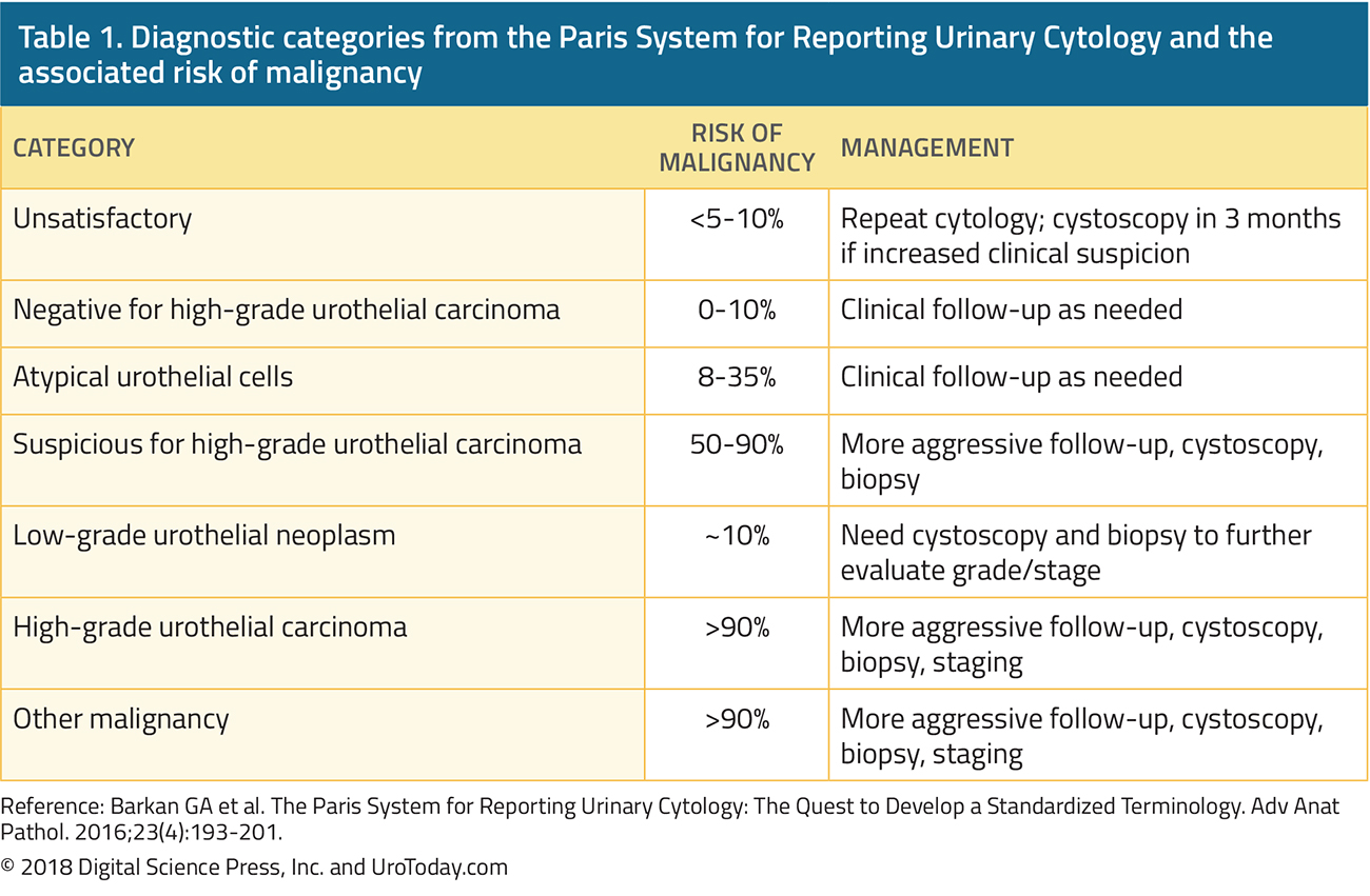
The urothelium is particularly well suited anatomically for assessment of potential biomarkers that can be obtained with little to no need for invasive procedures. Cells and cellular molecules (proteins, RNA, etc.) shed into the urine as it passes through the upper and lower urinary tract; they can be collected and purified from voided or catheterized specimens. The information gathered can then be used for screening, diagnosis, treatment response, and/or surveillance. The most well-known and widely used technique is urinary cytology, by which the cellular component of a urine specimen is microscopically assessed for features typically associated with high-grade malignancy (mitotic figures, condensed chromatin, enlarged nucleoli, etc.). Interpretation and reporting by cytopathologists has contributed to confusion surrounding urinary cytology with the use of terms like “suspicious, atypical, or indeterminate. The Paris Reporting System for Urinary Cytology has standardized the cytopathologic nomenclature while providing an estimated risk of malignancy based on associated literature (Table 1).15 While specificity has historically been very high (>99%), the poor sensitivity of urinary cytology, especially for papillary tumors (4-31%), make it far from ideal for either screening or surveillance.16-19 More contemporary data has been less robust, placing specificity at a more modest 82-88% and highlighting the need for more advanced markers.20
table-1-diagnosis-pathology-bladder-cancer@2x.jpg
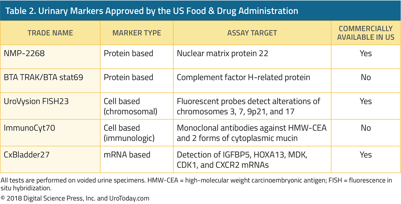
There are now five FDA-approved tests available (Table 2) in addition to many more potential biomarkers (i.e. DNA methylation, cell free DNA, histone modification) in various stages of development.21,22 The UroVysion test uses fluorescence in situ hybridization (FISH) to detect common chromosomal aberrations associated with bladder cancer and outperforms urinary cytology in terms of sensitivity, though this is almost entirely attributable to better detection of Ta tumors.23 The ImmunoCyt (uCyt+) assay was designed to complement cytology and increases the sensitivity to 59% for grade 1 tumors and up to 90% for grade 3 by using monoclonal antibodies directed against common urothelial surface markers.24 Nuclear matrix protein-22 (NMP-22) is involved in normal chromatin maintenance during mitosis but is greatly overexpressed in bladder carcinoma cells. Despite a reported sensitivity of 73% and specificity of 80%, the test has not gained widespread acceptance due to variability in accuracy between institutions and a high rate of false positive tests.25,26 The newest test to gain FDA-approval is the RNA based CxBladder test which measures the relative levels of 5 different mRNA transcripts within the urine (4 associated with malignancy and 1 with benign conditions) to produce an impressive combination of sensitivity and specificity at 82% and 85%, respectively.27,28 
table-2-diagnosis-pathology-bladder-cancer@2x.jpg
To date, none of the available data supports the use of urinary biomarkers as the sole method of bladder cancer detection, diagnosis, or follow-up, as stated by both the EAU and AUA in their respective NMIBC guidelines, however, they may offer useful information when assessing treatment response and during long-term surveillance as an adjunct to cystoscopy.7,8 Initial enthusiasm for these tests in the early to mid-2000s has waned, whereas use of urinary cytology has remained constant despite its shortcomings.29 The use of markers in prognostication and prediction of response to therapy is discussed in the next section on management of NMIBC.

Imaging

A complete diagnostic evaluation includes imaging of the entire urinary tract to assess for abnormalities of the urothelium normally out of view from cystoscopy. Upper tract urothelial tumors are uncommon, present in only 1.5% of patients with NMIBC, but certain features (multifocality, trigonal lesions, and/or CIS) raise this risk to more than 7%.30,31 The best combination of sensitivity (67-100%) and specificity (93-99%) is offered by computed tomographic urography (CTU) because of high soft tissue special resolution and contrast enhanced assessment of the urothelial surfaces and this has replaced intravenous urography in most centers in North America.32-35 Magnetic resonance imaging can be used as a substitute for CTU if the patient has an allergy to iodinated contrast or low GFR.36 In an effort to standardize MRI reporting and improve diagnostic accuracy, the multiparametric MRI based Vesical Imaging-Reporting and Data System (VI-RADS) was introduced in early 2018 with a 5 tiered system designed to predict likelihood of finding muscle invasion on TURBT, though it has not been validated in the clinical setting as of yet.37 Ultrasonography with retrograde pyelography is reserved for circumstances where both CT and MRI cannot be performed.

Pathology:

Urothelial carcinoma is the most common bladder cancer histology (~90%) diagnosed in the US, followed by squamous (2-5%), adenocarcinoma (2%), neuroendocrine (1%), and other rare tumors (<1%).38 The urothelium is the epithelial lining of the urinary tract and has a thickness in the bladder of approximately 5 to 7 cell layers overlying the lamina propria. Tumors that are confined to the bladder and do not invade the muscularis propria are considered non-muscle invasive bladder cancer (NMIBC) comprised of stages Ta, T1, and carcinoma in situ (CIS). Invasion of the muscularis propria – so called muscle invasive bladder cancer (MIBC,T2)“ represents an advanced stage with life threatening consequences requiring surgical management (i.e. radical cystectomy). 

Tumor Grading

Tumor grade is an important prognostic feature of bladder cancer but there is a lack of consensus internationally regarding the classification system. The extreme ends of the spectrum (highly aggressive and low malignant potential) are easy to identify but the middle ground has proved more elusive. In the World Health Organization 1973 grading system, there are 3 tiers of tumor grade (1, 2, and 3), though a majority of tumors end up as the intermediate grade 2 as a diagnosis of exclusion. The International Society of Urologic Pathologists (ISUP)/WHO 2004 grading system includes only high or low grade and exhibits better prognostic ability over the WHO 1973 system, at the expense of upward stage migration.39,40 Enrichment of the grade 3 group via inclusion of borderline grade 2 cases only leaves more indolent disease in the LG category and exposes those patients to overtreatment. The final version released as the ISUP/WHO 2004 grading system (now updated with minor revisions as the 2016 system) has been adopted throughout North America but the 1973 version is still in widespread use across Europe (Figure 1).41 
diagram-bladder-cancer@2x.jpg
Divergent Differentiation and Histologic Variants

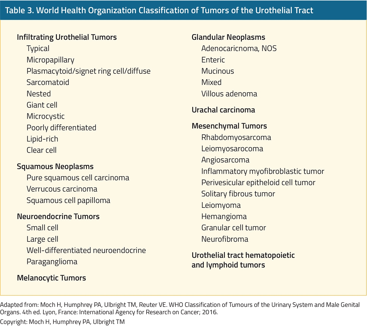
The ability of the urothelium to exhibit divergent differentiation is well known and may occur in a pure or mixed form (Table 3).42,43 When certain subtypes are present without elements of usual urothelial carcinoma, the tumor is referred to in terms of its pure histology (i.e. squamous cell carcinoma [SCC] of the bladder, adenocarcinoma of the bladder), in contrast to variant histology discussed below.41 Adenocarcinoma has a typical glandular (intestinal) appearance and tends to be more aggressive than UC.44 As such, complete early resection with radical cystectomy is advocated, even for T1 tumors, to achieve the best clinical outcomes.45 Pure SCC is more common in regions with endemic Bilhazrial infections and has been associated with a favorable clinical course, however, the non-infectious form is a distinct entity with a worse prognosis warranting similar management as adenocarcinoma.46 Small cell carcinoma is a neuroendocrine tumor with a very high propensity for distant spread at presentation and should be treated with upfront chemotherapy followed by surgery if free of detectable metastasis.47
table-3-diagnosis-pathology-bladder-cancer@2x.jpg
The impact of mixed histologic variants is less clear owing to multiple factors including low recognition historically among pathologists and tumor under-sampling during resection.48,49 In one study, repeat pathologic review of more than 1,200 bladder cancers diagnosed as pure UC between 1980 and 2005 found that 1/3rd actually contained a variant component.50 The sarcomatoid subtype is characterized by a mesenchymal and spindle-cell like appearance and exhibits a propensity for aggressive growth.51 These tumors present with extravesical invasion (T3-4) in about 1/3rd of cases.52 Micropapillary histologic architecture has been described in other malignancies and is typically associated with poor prognosis.53 Higher stage on presentation and increased likelihood for bladder invasion have been noted when even small regions of micropapillary differentiation are present (~10%), but the optimal treatment approach (neoadjuvant chemotherapy versus immediate RC) is still a topic for debate.54,55 Plasmacytoid variant is locally aggressive and frequently under-staged as evidenced by an 80% upstaging rate of clinical T1 to pathologic after cystectomy.56,57 The pattern of spread of plasmacytoid is particularly unusual for bladder cancer given its predilection for peritoneal implantation.58 The histologic appearance of nested subtype is similar to von Brunn nests, but unlike the benign nature of the latter, nested variant carries significant probability of muscle invasion (70%) and/or lymph node positivity (67%).59 Squamous and glandular differentiation are associated with a higher stage at initial diagnosis, however, clinical outcomes are no different than conventional urothelial carcinoma and standard treatment pathways should be sufficient.60,61 The AUA NMIBC guidelines direct the clinician to consider upfront radical cystectomy in T1 patients with any variant histology, citing the association of variant histology with a high rate of under-staging, however, the EAU guidelines limit their recommendation to only micropapillary histology.7,8,55,62

Molecular Classification

The accumulation of DNA damage necessary to produce bladder cancer requires several decades to occur, and as a result, MIBC exhibits a very high mutational burden and chromosomal instability. Concurrent genomic studies from several international research groups produced a range of intrinsic MIBC molecular subtypes with similar expressional profiles but different nomenclature. It is generally accepted that there are 2 major subtypes, luminal and basal, with better prognosis among the former. These tumors tend to be enriched for FGFR3 mutations associated with hyperproliferation and found at high frequency in non-invasive tumor. Despite the poor prognosis of the basal subtype, characterized by p53 mutations and alterations of DNA-damage repair pathways, these tumors are more responsive to platinum-based neoadjuvant chemotherapy than any other group.63 Low-grade tumors are genetically far more stable than MIBC, with highly conserved alterations in FGFR3 (79%), KDM6A (53%), and PIK3CA (52%).64,65 NMIBC has also been classified according to a molecular profiling schema, though it is less robust than what is available for MIBC. The major take-home point from this work thus far is the similarity between HG NMIBC and MIBC, pointing to a common pathway for progression.64,66,67

Published Date: April 16th, 2019

Written by: Justin T. Matulay, MD and Ashish Kamat, MD, MBBS
References: 1. Khadra MH, Pickard RS, Charlton M, et al. A prospective analysis of 1,930 patients with hematuria to evaluate current diagnostic practice. J Urol. 2000;163(2):524-527.
2. Mishriki SF, Nabi G, Cohen NP. Diagnosis of urologic malignancies in patients with asymptomatic dipstick hematuria: prospective study with 13 years' follow-up. Urology. 2008;71(1):13-16.
3. Divrik RT, Yildirim U, Zorlu F, et al. The effect of repeat transurethral resection on recurrence and progression rates in patients with T1 tumors of the bladder who received intravesical mitomycin: a prospective, randomized clinical trial. J Urol. 2006;175(5):1641-1644.
4. Gendy R, Delprado W, Brenner P, et al. Repeat transurethral resection for non-muscle-invasive bladder cancer: a contemporary series. BJU Int. 2016;117 Suppl 4:54-59.
5. Lazica DA, Roth S, Brandt AS, et al. Second transurethral resection after Ta high-grade bladder tumor: a 4.5-year period at a single university center. Urol Int. 2014;92(2):131-135.
6. Cumberbatch MGK, Foerster B, Catto JWF, et al. Repeat Transurethral Resection in Non-muscle-invasive Bladder Cancer: A Systematic Review. Eur Urol. 2018.
7. Babjuk M, Bohle A, Burger M, et al. EAU Guidelines on Non-Muscle-invasive Urothelial Carcinoma of the Bladder: Update 2016. Eur Urol. 2017;71(3):447-461.
8. Chang SS, Boorjian SA, Chou R, et al. Diagnosis and Treatment of Non-Muscle Invasive Bladder Cancer: AUA/SUO Guideline. J Urol. 2016;196(4):1021-1029.
9. Chang TC, Marcq G, Kiss B, et al. Image-Guided Transurethral Resection of Bladder Tumors - Current Practice and Future Outlooks. Bladder Cancer. 2017;3(3):149-159.
10. O'Brien T, Ray E, Chatterton K, et al. Prospective randomized trial of hexylaminolevulinate photodynamic-assisted transurethral resection of bladder tumour (TURBT) plus single-shot intravesical mitomycin C vs conventional white-light TURBT plus mitomycin C in newly presenting non-muscle-invasive bladder cancer. BJU Int. 2013;112(8):1096-1104.
11. Schumacher MC, Holmang S, Davidsson T, et al. Transurethral resection of non-muscle-invasive bladder transitional cell cancers with or without 5-aminolevulinic Acid under visible and fluorescent light: results of a prospective, randomised, multicentre study. Eur Urol. 2010;57(2):293-299.
12. Yuan H, Qiu J, Liu L, et al. Therapeutic outcome of fluorescence cystoscopy guided transurethral resection in patients with non-muscle invasive bladder cancer: a meta-analysis of randomized controlled trials. PLoS One. 2013;8(9):e74142.
13. Herr HH. Narrow band imaging cystoscopy. Urol Oncol. 2011;29(4):353-357.
14. Kang W, Cui Z, Chen Q, et al. Narrow band imaging-assisted transurethral resection reduces the recurrence risk of non-muscle invasive bladder cancer: A systematic review and meta-analysis. Oncotarget. 2017;8(14):23880-23890.
15. Barkan GA, Wojcik EM, Nayar R, et al. The Paris System for Reporting Urinary Cytology: The Quest to Develop a Standardized Terminology. Adv Anat Pathol. 2016;23(4):193-201.
16. Lotan Y, Roehrborn CG. Sensitivity and specificity of commonly available bladder tumor markers versus cytology: results of a comprehensive literature review and meta-analyses. Urology. 2003;61(1):109-118; discussion 118.
17. Schmitz-Drager BJ, Droller M, Lokeshwar VB, et al. Molecular markers for bladder cancer screening, early diagnosis, and surveillance: the WHO/ICUD consensus. Urol Int. 2015;94(1):1-24.
18. Xylinas E, Kluth LA, Rieken M, et al. Urine markers for detection and surveillance of bladder cancer. Urol Oncol. 2014;32(3):222-229.
19. Zuiverloon TCM, de Jong FC, Theodorescu D. Clinical Decision Making in Surveillance of Non-Muscle-Invasive Bladder Cancer: The Evolving Roles of Urinary Cytology and Molecular Markers. Oncology (Williston Park). 2017;31(12):855-862.
20. Yafi FA, Brimo F, Auger M, et al. Is the performance of urinary cytology as high as reported historically? A contemporary analysis in the detection and surveillance of bladder cancer. Urol Oncol. 2014;32(1):27 e21-26.
21. Chou R, Gore JL, Buckley D, et al. Urinary Biomarkers for Diagnosis of Bladder Cancer: A Systematic Review and Meta-analysis. Ann Intern Med. 2015;163(12):922-931.
22. Santoni G, Morelli MB, Amantini C, et al. Urinary Markers in Bladder Cancer: An Update. Front Oncol. 2018;8:362.
23. Hajdinjak T. UroVysion FISH test for detecting urothelial cancers: meta-analysis of diagnostic accuracy and comparison with urinary cytology testing. Urol Oncol. 2008;26(6):646-651.
24. Comploj E, Mian C, Ambrosini-Spaltro A, et al. uCyt+/ImmunoCyt and cytology in the detection of urothelial carcinoma: an update on 7422 analyses. Cancer Cytopathol. 2013;121(7):392-397.
25. Shariat SF, Marberger MJ, Lotan Y, et al. Variability in the performance of nuclear matrix protein 22 for the detection of bladder cancer. J Urol. 2006;176(3):919-926; discussion 926.
26. Poulakis V, Witzsch U, De Vries R, et al. A comparison of urinary nuclear matrix protein-22 and bladder tumour antigen tests with voided urinary cytology in detecting and following bladder cancer: the prognostic value of false-positive results. BJU Int. 2001;88(7):692-701.
27. Kavalieris L, O'Sullivan PJ, Suttie JM, et al. A segregation index combining phenotypic (clinical characteristics) and genotypic (gene expression) biomarkers from a urine sample to triage out patients presenting with hematuria who have a low probability of urothelial carcinoma. BMC urology. 2015;15:23.
28. O'Sullivan P, Sharples K, Dalphin M, et al. A multigene urine test for the detection and stratification of bladder cancer in patients presenting with hematuria. J Urol. 2012;188(3):741-747.
29. Narayan VM, Adejoro O, Schwartz I, et al. The Prevalence and Impact of Urinary Marker Testing in Patients with Bladder Cancer. J Urol. 2018;199(1):74-80.
30. Palou J, Rodriguez-Rubio F, Huguet J, et al. Multivariate analysis of clinical parameters of synchronous primary superficial bladder cancer and upper urinary tract tumor. J Urol. 2005;174(3):859-861; discussion 861.
31. Millan-Rodriguez F, Chechile-Toniolo G, Salvador-Bayarri J, et al. Upper urinary tract tumors after primary superficial bladder tumors: prognostic factors and risk groups. J Urol. 2000;164(4):1183-1187.
32. Chlapoutakis K, Theocharopoulos N, Yarmenitis S, et al. Performance of computed tomographic urography in diagnosis of upper urinary tract urothelial carcinoma, in patients presenting with hematuria: Systematic review and meta-analysis. Eur J Radiol. 2010;73(2):334-338.
33. Cowan NC, Turney BW, Taylor NJ, et al. Multidetector computed tomography urography for diagnosing upper urinary tract urothelial tumour. BJU Int. 2007;99(6):1363-1370.
34. Froemming A, Potretzke T, Takahashi N, et al. Upper tract urothelial cancer. Eur J Radiol. 2018;98:50-60.
35. Roupret M, Babjuk M, Comperat E, et al. European Association of Urology Guidelines on Upper Urinary Tract Urothelial Carcinoma: 2017 Update. Eur Urol. 2018;73(1):111-122.
36. Takahashi N, Glockner JF, Hartman RP, et al. Gadolinium enhanced magnetic resonance urography for upper urinary tract malignancy. J Urol. 2010;183(4):1330-1365.
37. Panebianco V, Narumi Y, Altun E, et al. Multiparametric Magnetic Resonance Imaging for Bladder Cancer: Development of VI-RADS (Vesical Imaging-Reporting And Data System). Eur Urol. 2018;74(3):294-306.
38. Hansel DE, Amin MB, Comperat E, et al. A contemporary update on pathology standards for bladder cancer: transurethral resection and radical cystectomy specimens. Eur Urol. 2013;63(2):321-332.
39. Cao D, Vollmer RT, Luly J, et al. Comparison of 2004 and 1973 World Health Organization Grading Systems and Their Relationship to Pathologic Staging for Predicting Long-term Prognosis in Patients With Urothelial Carcinoma. Urology. 2010;76(3):593-599.
40. Lokeshwar SD, Ruiz-Cordero R, Hupe MC, et al. Impact of 2004 ISUP/WHO classification on bladder cancer grading. World Journal of Urology. 2015;33(12):1929-1936.
41. Humphrey PA, Moch H, Cubilla AL, et al. The 2016 WHO Classification of Tumours of the Urinary System and Male Genital Organs-Part B: Prostate and Bladder Tumours. Eur Urol. 2016;70(1):106-119.
42. Lopez-Beltran A, Cheng L. Histologic variants of urothelial carcinoma: differential diagnosis and clinical implications. Hum Pathol. 2006;37(11):1371-1388.
43. Shanks JH, Iczkowski KA. Divergent differentiation in urothelial carcinoma and other bladder cancer subtypes with selected mimics. Histopathology. 2009;54(7):885-900.
44. Wright JL, Porter MP, Li CI, et al. Differences in survival among patients with urachal and nonurachal adenocarcinomas of the bladder. Cancer. 2006;107(4):721-728.
45. Zaghloul MS, Nouh A, Nazmy M, et al. Long-term results of primary adenocarcinoma of the urinary bladder: a report on 192 patients. Urol Oncol. 2006;24(1):13-20.
46. Ehdaie B, Maschino A, Shariat SF, et al. Comparative outcomes of pure squamous cell carcinoma and urothelial carcinoma with squamous differentiation in patients treated with radical cystectomy. J Urol. 2012;187(1):74-79.
47. Lynch SP, Shen Y, Kamat A, et al. Neoadjuvant chemotherapy in small cell urothelial cancer improves pathologic downstaging and long-term outcomes: results from a retrospective study at the MD Anderson Cancer Center. Eur Urol. 2013;64(2):307-313.
48. Willis D, Kamat AM. Nonurothelial bladder cancer and rare variant histologies. Hematol Oncol Clin North Am. 2015;29(2):237-252, viii.
49. Shah RB, Montgomery JS, Montie JE, et al. Variant (divergent) histologic differentiation in urothelial carcinoma is under-recognized in community practice: Impact of mandatory central pathology review at a large referral hospital. Urol Oncol. 2013;31(8):1650-1655.
50. Linder BJ, Boorjian SA, Cheville JC, et al. The impact of histological reclassification during pathology re-review--evidence of a Will Rogers effect in bladder cancer? J Urol. 2013;190(5):1692-1696.
51. Moch H, Humphrey PA, Ulbright TM, et al. WHO Classification of Tumours of the Urinary System and Male Genital Organs. 4th ed. Lyon, France: International Agency for Research on Cancer; 2016.
52. Sui W, Matulay JT, Onyeji IC, et al. Contemporary treatment patterns and outcomes of sarcomatoid bladder cancer. World J Urol. 2017;35(7):1055-1061.
53. Sui W, Matulay JT, James MB, et al. Micropapillary Bladder Cancer: Insights from the National Cancer Database. Bladder Cancer. 2016;2(4):415-423.
54. Kamat AM, Dinney CP, Gee JR, et al. Micropapillary bladder cancer: a review of the University of Texas M. D. Anderson Cancer Center experience with 100 consecutive patients. Cancer. 2007;110(1):62-67.
55. Meeks JJ, Taylor JM, Matsushita K, et al. Pathological response to neoadjuvant chemotherapy for muscle-invasive micropapillary bladder cancer. BJU Int. 2013;111(8):E325-330.
56. Kaimakliotis HZ, Monn MF, Cary KC, et al. Plasmacytoid variant urothelial bladder cancer: is it time to update the treatment paradigm? Urol Oncol. 2014;32(6):833-838.
57. Keck B, Wach S, Stoehr R, et al. Plasmacytoid variant of bladder cancer defines patients with poor prognosis if treated with cystectomy and adjuvant cisplatin-based chemotherapy. BMC Cancer. 2013;13:71.
58. Ricardo-Gonzalez RR, Nguyen M, Gokden N, et al. Plasmacytoid carcinoma of the bladder: a urothelial carcinoma variant with a predilection for intraperitoneal spread. J Urol. 2012;187(3):852-855.
59. Wasco MJ, Daignault S, Bradley D, et al. Nested variant of urothelial carcinoma: a clinicopathologic and immunohistochemical study of 30 pure and mixed cases. Hum Pathol. 2010;41(2):163-171.
60. Amin MB. Histological variants of urothelial carcinoma: diagnostic, therapeutic and prognostic implications. Mod Pathol. 2009;22 Suppl 2:S96-S118.
61. Mitra AP, Bartsch CC, Bartsch G, Jr., et al. Does presence of squamous and glandular differentiation in urothelial carcinoma of the bladder at cystectomy portend poor prognosis? An intensive case-control analysis. Urol Oncol. 2014;32(2):117-127.
62. Vetterlein MW, Wankowicz SAM, Seisen T, et al. Neoadjuvant chemotherapy prior to radical cystectomy for muscle-invasive bladder cancer with variant histology. Cancer. 2017;123(22):4346-4355.
63. Seiler R, Ashab HA, Erho N, et al. Impact of Molecular Subtypes in Muscle-invasive Bladder Cancer on Predicting Response and Survival after Neoadjuvant Chemotherapy. Eur Urol. 2017.
64. Hurst CD, Alder O, Platt FM, et al. Genomic Subtypes of Non-invasive Bladder Cancer with Distinct Metabolic Profile and Female Gender Bias in KDM6A Mutation Frequency. Cancer Cell. 2017;32(5):701-715 e707
65. Robertson AG, Kim J, Al-Ahmadie H, et al. Comprehensive Molecular Characterization of Muscle-Invasive Bladder Cancer. Cell. 2017;171(3):540-556 e525.
66. Hedegaard J, Lamy P, Nordentoft I, et al. Comprehensive Transcriptional Analysis of Early-Stage Urothelial Carcinoma. Cancer Cell. 2016;30(1):27-42.
67. Pietzak EJ, Bagrodia A, Cha EK, et al. Next-generation Sequencing of Nonmuscle Invasive Bladder Cancer Reveals Potential Biomarkers and Rational Therapeutic Targets. Eur Urol. 2017;72(6):952-959.
68. Soloway MS, Briggman V, Carpinito GA, et al. Use of a new tumor marker, urinary NMP22, in the detection of occult or rapidly recurring transitional cell carcinoma of the urinary tract following surgical treatment. J Urol. 1996;156(2 Pt 1):363-367.
69. Irani J, Desgrandchamps F, Millet C, et al. BTA stat and BTA TRAK: A comparative evaluation of urine testing for the diagnosis of transitional cell carcinoma of the bladder. Eur Urol. 1999;35(2):89-92.
70. Fradet Y, Lockhard C. Performance characteristics of a new monoclonal antibody test for bladder cancer: ImmunoCyt trade mark. Can J Urol. 1997;4(3):400-405.
71. Mostofi FK, Sobin LH, Torloni H. Histological typing of urinary bladder tumours. Geneva, Switerland: World Health Organization; 1973.
72. Eble JN, Sauter G, Epstein JI, et al. Pathology and Genetics of Tumors of the Urinary System and Male Genital Organs. Lyon: IARC Press; 2004.