Figure 1 – 3D Models:
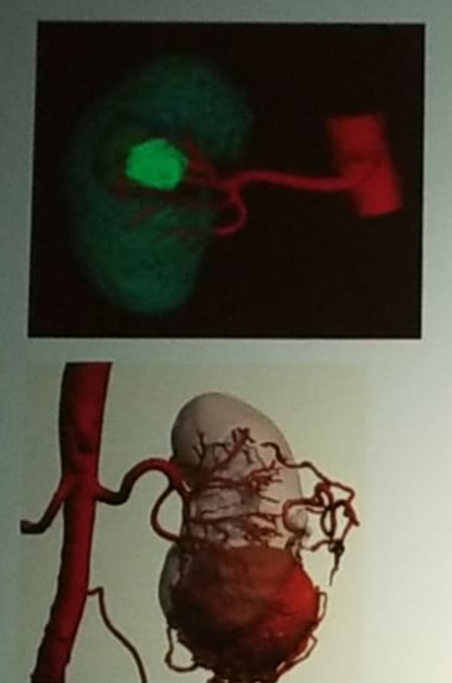
The creation of an accurate 3D understanding of the anatomy, based on over 2000 images from a standard CT or MRI scan is challenging. Studies have been done to test how accurate is the surgeon’s understanding of the anatomy after reviewing the CT or MRI scans. Urologists were asked to first review the CT or MRI scans of a kidney with a tumor, and then they were tasked with identifying the precise location of the tumor on a 3D kidney model. Surprisingly, the urologists were correct only 25% of the time.
The process of familiarizing oneself with the software that produces the 3D images was initially daunting and shown to be very time-consuming. Generalizing one’s first model required 8-10 hours initially, and achieving a master level required making at least 3-5 models.
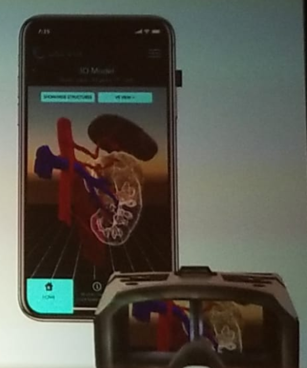
Recently, a healthcare startup, called CEEVRA, created an application, which can be downloaded to a smartphone and can create beautiful, detailed 3D models created from existing CTs and MRIs. It can be viewed from the surgeon’s smartphone, and optionally, through a basic virtual reality headset (Figure 2). It is a full cloud-based software service, and is FDA cleared for both preoperative planning and intraoperative display and is also HIPAA compliant. A multicenter randomized controlled clinical study for robotic partial nephrectomy was completed just recently. The study’s endpoints included operative time, estimated blood loss, clamp time, and length of hospital stay. The study’s results are pending publication. According to Dr. Herrell, the preoperative plan was modified in a third of the cases resulting from the usage of this application.
Figure 2 – CEEVRA Application which can be Downloaded to the Surgeon’s Smartphone:
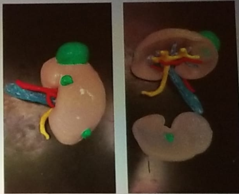
Dr. Herrel concluded his discussion, mentioning the role of 3D printed models. Currently, there are several 3d printed models which are being investigated (Figure 3). The price of these 3D printers has significantly dropped, and it is possible to use them to print a model of segmented CT scan, demonstrating key anatomical features, including vessels, collecting system, and tumors and margins. It enables manual manipulation and assessment, but the disadvantages include the fact that the printed model is only as good as the segmentation CT scan. Furthermore, the fact that it is typically printed in hard plastic does not enable surgical practice.
Figure 3- Printed 3D Models:
Presented by: S. Duke Herrell, MD. Professor, Department of Urology, Vanderbilt University, Nashville, Tennessee
Written By: Hanan Goldberg, MD, Urologic Oncology Fellow (SUO), University of Toronto, Princess Margaret Cancer Centre @GoldbergHanan at 2019 3rd Annual North American Robotic Urology Symposium (NARUS), February 8-9, 2018 - Las Vegas, Nevada, United States