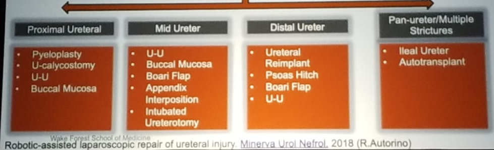
When facing a ureteral stricture, it is important to make sure that appropriate imaging studies are available, including CT urogram, or MR urogram, and renal scan or antegrade nephrostogram, if available. The imaging should be used to delineate the etiology, location, length of ureteral stricture, and degree of fibrosis. Depending on the location of the ureteral stricture, various therapeutic options are available, as can be seen in figure 1.
Figure 1- Possible Therapeutic Options for Ureteral Strictures:
Boari flap is a useful option when the diseased segment of the ureter is too long, or ureteral mobility is too limited to perform a primary ureteroureterostomy. Boari flaps can be created to bridge a 10- to 15-cm ureteral defect. If needed, spiral bladder flaps can be constructed to reach the renal pelvis in some circumstances.
The robot is ideal for creating the Boari flap according to Dr. Hemal. It can be used to create the Boari flap up to the level of L3-L4. A cystogram needs to be performed to outline bladder contour and determine its capacity. The robotic ports placement for the creation of a Boari flap is shown in Figure 2.
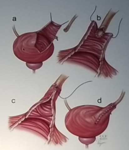
The actual technique entails (Figure 3):
1. Identification of the ureter at the bifurcation of the common iliac artery or above and mobilization of it caudally until you can identify the stricture site.
2. Mobilization of the bladder as distally as possible on both sides
3. The ratio between the length and width of the bladder flap is 2:1 (classic teaching in open surgery was 5:1).
4. Performing a Psoas hitch is required in every case. A psoas hitch entails mobilization of the bladder with fixation above the iliac vessels to guarantee a tension-free ureteric anastomosis.
5. The ureter is spatulated, and a sub-mucosal tunnel is created at the cranial aspect of the Boari’s flap and the ureter is carefully tunneled through the sub-mucosal tunnel into the bladder.
6. The ureter is then sutured to the mucosa of the flap using interrupted sutures.
7. Tubularization of the bladder flap takes place with continuous sutures placed in two layers.
8. The bladder opening is then closed in two layers using continuous sutures.
9. The watertight suturing of the anastomosis and bladder is finally tested.
Figure 2 – Port placement for Boari Flap:
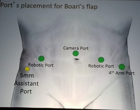
Figure 3 – Boari Flap Technique:
Important principles that need to be addressed during this procedure include a tension-free anastomosis, spatulation of the ureter, water-tight anastomosis, keeping the ureter vascularized, and performing extravesical reimplantation. It is critical to keep in mind and practice these issues when attempting to perform the Boari flap:
1.Removal of an obstructed/diseased /unhealthy ureteral segment
2.Proximal ureterolysis with the maintenance of blood supply
3.Mobilization of the bladder
4. Tension-free anastomosis with 4-0 or 5-0 sutures
5. The psoas hitch should be performed with ethibond or PDS
6. Ensuring adequate blood supply to the anastomotic area.
Dr. Homel concluded this technical presentation with some important points. Most ureteral pathologies including complex upper ureteric reconstruction can be performed with the robot. Robotic Boari flap can be used for unilateral and bilateral distal ureteral strictures. Lastly, the experience of the surgical team is paramount in handling these diverse ureteric aetiologies, requiring different operative procedures.
Presented by: Ashok K Hemal, MD, Wake Forrest Baptist Health Urology, Winston-Salem, North Carolina
Written By: Hanan Goldberg, MD, Urologic Oncology Fellow (SUO), University of Toronto, Princess Margaret Cancer Centre @GoldbergHanan at 2019 3rd Annual North American Robotic Urology Symposium (NARUS), February 8-9, 2018 - Las Vegas, Nevada, United States