Kidney Cancer Today
The Ongoing Evolution of a Field: Advances In First Line Therapy For Metastatic Clear Cell Renal Cell Carcinoma
Introduction
Cancers of the kidney and renal pelvis (when considered in aggregate despite different histology) represent the 6th most common newly diagnosed tumors in men and 8th most common in women in the United States in 20201, representing an estimated 73,750 new diagnoses and 14,830 deaths. The vast majority of these cancers will be renal parenchymal tumors with renal cell carcinoma (RCC) comprising the large majority with clear cell renal cell carcinoma (ccRCC) is the most common histologic subtype of renal cell carcinoma. Due to its prevalence, the vast majority of advances in systemic therapies for RCC have been made for patients with ccRCC. However, there have been important recent advances in treatment for patients with non-clear cell renal cell carcinomas (nccRCC) as well in recent years.
Despite ongoing stage migration as a result of widespread use of axial abdominal imaging for non-specific abdominal complaints2, a large proportion (up to 35%) of patients present with advanced disease, including metastases3. Historically, metastatic RCC has been early uniformly fatal, with 10-year survival rates less than 5%4. However, there has been transformational change in this disease space over the past fifteen years and, with newer immunotherapy-based approaches, the potential for long-term cure is something that may be considered. Certainly, a significantly longer natural history is feasible given available therapeutic options.
The Historical and Near Past
The immunologically active nature of RCC has been recognized for many years and, as a result, modulators of the immune system were among the first therapeutic targets for advanced ccRCC: interferon-alfa and interleukin-2 were among the only available treatment options prior to 2005. However, despite a response rate between 10 to 15%5, even among patients treated at a center of excellence, median overall survival was only 30 months in favorable risk patients, 14 months in intermediate risk patients and 5 months in poor risk patients6. Interleukin-2 had similar response rates to interferon-based therapies (~15 to 20%)7, but distinctly had evidence of durable complete responses in approximately 7 to 9% of patients8. This observation led to the U.S. Food and Drug Administration (FDA) approval of high-dose IL-2 in 1992. However, IL-2 is associated with significant toxicity which has limited its widespread use.
The more recent past includes the “targeted therapy” era which began with the introduction of sorafenib in 2005 followed by sunitinib in 2006 and temsirolimus in 2007, along with a number of other agents in the years that followed.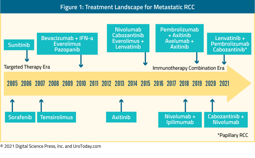
These treatments were developed based on work into the molecular biology underlying ccRCC through targeting of the vascular endothelial growth factor (VEGF) pathway and mammalian target of rapamycin (mTOR). This pathway plays a key role in regulating HIF-α, thus modulating the pathway between abnormalities in VHF and proliferation.
While no longer used as monotherapy in the first-line setting, bevacizumab, a humanized monoclonal antibody against VEGF-A, was the first inhibitor of the VEGF pathway used in clinical trials. In head-to-head trials against interferon-alfa, the addition of bevacizumab to interferon resulted in significant improvements in response rate and progression-free survival9,10.
In contrast, tyrosine-kinase inhibitors (TKIs) quickly became standard of care, used as first-line monotherapy. For nearly 15 years, sunitinib was the standard of care, and as such, it has formed the control comparison for testing of newer approaches. As with bevacizumab, TKIs also target the VEGF pathway, through inhibition of a combination of VEGFR-2, PDGFR-β, raf-1 c-Kit, and Flt3 (sunitinib and sorafenib). As alluded to above, sorafenib was one the first molecularly targeted agents clinically available, in 2006, based on demonstrated biologic activity in ccRCC. However, despite FDA approval, sorafenib was quickly supplanted by sunitinib as a first-line VEGF inhibitor. Sunitinib was first tested among patients who had previously received cytokine therapy and then, in a pivotal phase III trial, demonstrated superiority (both in terms of progression free survival and quality of life) in a head-to-head comparison with interferon-α11. Since the approval of sunitinib and sorafenib, there has been development and subsequent approval of many other tyrosine kinase inhibitors. For the most part, the goal of these agents has been to reduce the toxicity of VEGF inhibitors while retaining oncologic efficacy. Comparative data of pazopanib and sunitinib have demonstrated non-inferior oncologic outcomes with decreased toxicity among patients receiving pazopanib12. Axitinib was evaluated first as second-line therapy13 and then in the first-line setting compared to sorafenib14. Finally, tivozanib has been compared to sorafenib among patients who had not previously received VEGF or mTOR-targeting therapies. While this study demonstrated tivozanib’s activity, it was not FDA approved and it therefore not used.
Most recently, cabozantinib, a multikinase inhibitor (acting on tyrosine kinases including MET, VEGF receptors), and TAM family of kinases (TYRO3, MER, and AXL), has been approved for the first-line treatment of mRCC based on the phase II CABOSUN trial. In the initial report of this study, cabozantinib demonstrated significantly improved progression free survival (HR 0.66, 95% CI 0.46 to 0.95), compared to sunitinib in the first line treatment of patients with intermediate or poor risk mRCC15. In an updated analysis utilizing independent PFS review, comparable PFS results were observed (HR 0.48, 95% CI 0.31 to 0.74)16. However, this trial has yet to demonstrate an overall survival benefit to cabozantinib compared to sunitinib (HR 0.80, 95% CI 0.53 to 1.21).
Mammalian target of rapamycin (mTOR) inhibitors were developed in parallel to VEGF inhibitors. Unlike TKIs, for the most part, these agents have not been used in first-line therapy, though temsirolimus has been used in patients with poor-risk disease based on a comparison or temsirolimus, interferon, and the combination in 626 patients with pre-defined poor risk metastatic RCC who had not previously received systemic therapy17. Patients who received temsirolimus had significantly improved overall survival compared to those receiving interferon-alfa (HR 0.73, 95% CI 0.58 to 0.92).
In addition to the recent, though now well-established, role of immunotherapy in patients with mRCC, there remains ongoing interest in the development of targeted therapies based on our understanding of mRCC biology. Notably, on the basis of an understanding of ccRCC carcinogenesis, the potent, selective, small molecular HIF-2α inhibitor belzutifan (MK-6482) was granted priority review by the FDA for patients with von Hippel-Lindau (VHL) associated RCC. This approval was provided based on the phase II Study-004 trial among patients with renal tumors not requiring surgical intervention (NCT03401788). In data presented at ASCO-GU 2021, treatment with belzutifan was associated with an overall response rate of 36.1% (95% confidence interval 24.2-49.4%). MK-6482 also demonstrated benefits in non-RCC tumors including pancreatic lesions and central nervous systemic hemangioblastomas. This novel therapy was relatively well tolerated with 13% experiencing grade 3 treatment-related adverse events and none experiencing grade 4 or 5 treatment-related adverse events. While this approval was based on treatment in patients with renal masses not requiring surgical intervention, there are ongoing phase III trials of belzutifan both as monotherapy and in combination regimes as first-line treatment for advanced ccRCC.
The Return of Immunotherapy for Advanced RCC
While the cytokine era faded with the introduction of targeted therapies, the immunologic basis for mRCC treatment re-emerged around 2015 with the use of nivolumab monotherapy for patients who had previously received systemic therapy.
Published now more than 3 years ago, CheckMate 214 was the first study to demonstrate a benefit for immune checkpoint inhibitors in the first-line treatment of mRCC, showing an overall survival (OS) benefit for first-line nivolumab + ipilimumab vs sunitinib18. This trial randomized 1096 patients to the combination immunotherapy approach of nivolumab + ipilimumab (550 patients) or sunitinib (546 patients). Most patients had intermediate or poor risk disease (n=847). OS was significantly improved in the overall patient population; however, stratified analyses provide more nuanced results with benefits restricted to those with intermediate or poor-risk RCC while, in patients with favorable risk disease, progression-free survival and overall response rate were higher among patients who received sunitinib.
Since the initial publication, there have been a number of follow-up and subgroup analyses from CheckMate 214. Long-term follow-up among patients with at least four years of follow-up was reported at ESMO 2020 by Dr. Albiges. Among these patients, in the intention-to-treat population, results were very similar to the initial analysis previously published with the combined nivolumab + ipilimumab approach continuing to demonstrate superiority (HR 0.69, 95% CI 0.59 to 0.81). In sub-groups defined according to IMDC criteria, those with intermediate or poor risk had improved survival with nivolumab/ipilimumab (HR 0.65, 95% CI 0.54 to 0.78) while there continued to be no appreciable difference between treatment approaches among those with favorable risk disease (HR 0.93, 95% CI 0.62 to 1.40). Presented at the same meeting, Dr. Regan and colleagues used these long-term follow-up data to assess a novel outcome metric, treatment-free survival with and without toxicity. The rationale for this approach is that conventional measures (OS, rPFS, etc) may not fully capture the effects on immuno-oncology (IO) approaches, particularly patients may have long periods of disease control without subsequent anticancer therapy following discontinuation of IO regimes. Thus, the authors defined treatment free survival (TFS), as the time between protocol therapy cessation and subsequent systemic therapy or death. They stratified this as TFS with or without toxicity by counting the number of days with ≥1 grade ≥3 treatment-related adverse events reported. As of 42-months of follow-up, 56% of patients randomized to nivolumab + ipilimumab and 47% of those randomized to sunitinib were alive with 13% and 7%, respectively, remaining on their original therapy. A further 31% of patients randomized to nivolumab + ipilimumab and 12% of those randomized to sunitinib were surviving free of subsequent, second line therapy. 42-month restricted TFS was higher for patients randomized to nivolumab + ipilimumab (7.8 months) than those randomized to sunitinib (3.3 months). Toxicity-free TFS was 7.1 months and 3.0 months, respectively. In each case, the 95% confidence interval of the difference in median TFS excluded unity demonstrating that these are significant differences. Unlike the differences in PFS and OS which appear to be restricted to patients with intermediate and poor risk disease, Dr. Regan and colleagues showed that the benefits in TFS were dramatic in patients with both IMBC intermediate and poor risk disease (median TFS 6.9 vs 3.1 months) and favourable risk disease (median TFS 11.0 vs 3.7 months).
One of the final important subgroup analyses comes from Dr. Escudier and colleagues who demonstrated that the objective response rate was stable across increasing numbers of IMDC risk factors (from zero to 6) for those who received nivolumab and ipilimumab, while the ORR in patients treated with sunitinib decreased with an increasing number of IMDC risk factors19.
The BIONIKK trial, an open-label, phase II biomarker-driven randomized trial, was also presented as ESMO 2020. This trial relied upon previous analyses which demonstrated that immune and angiogenic signatures can allow for the differentiation of four groups of patients (ccrcc1-4) with immune and angiogenic high/low features, which could allow better identification of responders to either nivolumab, nivolumab + ipilimumab or TKI. ccrcc1 “immune-low” and ccrcc4 “immune-high” tumors have been associated with the poorest outcomes, whereas ccrcc2 “angio-high” and ccrcc3 “normal-like” tumors have been associated with the best outcomes. In this biomarker driven trial, patients with ccrcc1 and ccrcc4 signatures were randomized to nivolumab versus nivolumab + ipilimumab, whereas those with ccrcc2 and ccrcc3 signatures were randomized to receive nivolumab + ipilimumab versus TKI. As a phase II trial, the primary endpoint for this study was objective response rate (ORR, RECIST1.1) per treatment and group. Secondary endpoints included PFS, OS, and tolerability. 202 patients were randomized of a targeted 187. Among patients with the ccrcc1 signature, objective response rates were higher among those who received combination therapy with nivolumab + ipilimumab (39.4%; 6.1% complete response rate) than those who received nivolumab alone (20.7%; 0% complete response rate) whereas among those with a ccrcc4 signature, objective response rates were 50.3% in those receiving the combination approach (11.8% complete response rate) as compared to 50% in those receiving nivolumab alone (7.1%). Median progression free survival among patients with the ccrcc1 signature was 8.0 months in those receiving nivolumab + ipilimumab and 4.6 months among those receiving nivolumab alone. In the ccrcc4 group, median progression-free survival was 12.2 months in the combination arm and 7.8 months in the nivolumab monotherapy arm. In patients with the ccrcc2 signature, objective response rates were 48.3% in the nivolumab + ipilimumab arm (13.8% complete response rate) and 53.8% in the TKI arm (0% complete response rate) whereas among patients with the ccrcc3 signature, 25% receiving nivolumab + ipilimumab had objective responses (0% complete response rate) and 0% receiving TKI had objective response. These are the first randomized data based on molecular risk group assessment to guide first-line therapy in metastatic ccRCC. In particular, among patients with the ccrcc4 signature, use of combination therapy may not be required and thus ipilimumab may be spared.
In terms of first line therapy, immunotherapy approaches have predominately focused on combination therapy approaches. However, in the past month, data regarding the use of pembrolizumab monotherapy has emerged20. This phase II single-arm study demonstrated an objective response rate of 36.4% among 110 enrolled patients with a median progression-free survival of 7.1 months (95% CI 5.6 to 11.0 months). Clearly, compared to the data highlighted both above from CheckMate214 and in the sections that follow, these results are inferior to combination therapy.
Combination Approaches: Targeted Therapy and Immunotherapy
Combination therapy has been well established in the treatment of advanced RCC, including the use of interferon-alfa and bevacizumab9,10. Following the data from CheckMate 214 demonstrating the role for immune checkpoint blockade in advanced RCC, data began to emerge on the combination of targeted therapies with checkpoint inhibitors.
The first of these studies was IMmotion151, first presented at GU ASCO 2018 and subsequently published, which compared first-line atezolizumab + bevacizumab vs sunitinib among 915 patients with previously untreated metastatic RCC21. The combined approach demonstrated a significant benefit in progression-free survival (11.2 months versus 7.7 months; HR 0.74, 95% CI 0.57 to 0.96) among the whole cohort of patients and had lower rates of significant (grade 3-4) adverse events (40% vs 54%).
Subsequently, further combination approaches have been approved on the basis of published phase III trials, including pembrolizumab + axitinib (KEYNOTE-426) and avelumab + axitinib (JAVELIN Renal 101). Additionally, the recent presentation and publication of CheckMate-9ER and CLEAR have added the combination of nivolumab + cabozantinib and lenvatinib + pembrolizumab, respectively, to the armamentarium of first line mRCC treatment.
In KEYNOTE-426, 861 patients with metastatic clear cell RCC, predominately with intermediate or poor risk disease, who had not previously received systemic therapy were randomized to pembrolizumab + axitinib or sunitinib and followed for the co-primary endpoints of overall survival and progression free survival22. While median OS was not reached, patients who received pembrolizumab + axitinib had improved OS (HR 0.53, 95% CI 0.38 to 0.74) and progression free survival (HR 0.69, 95% CI 0.57 to 0.84), as well as overall response rate. These results were consistent across subgroups of demographic characteristics, IMDC risk categories, and PD-L1 expression level. Grade 3 to 5 adverse events were somewhat more common among patients getting pembrolizumab and axitinib, though rates of discontinuation were lower.
Similarly, JAVELIN Renal 101 randomized 886 patients to avelumab + axitinib or sunitinib23. Again, the preponderance of patients had IMDC intermediate or poor risk disease. In this analysis the primary endpoints were PFS and OS in patients with PD-L1 positive tumors. Notably, 560 of the 886 patients had PD-L1 positive tumors. Among the PD-L1 positive subgroup, progression free survival (HR 0.61, 95% CI 0.47 to 0.79) was improved in patients receiving avelumab + axitinib compared to sunitinib while OS did not significantly differ (HR 0.82, 95% CI 0.53 to 1.28). In the overall study population, progression-free survival was similarly improved, as compared to the PD-L1 positive population (HR 0.69, 95% CI 0.56 to 0.84).
Third, in data initially presented at ESMO 2020 and published in February 2021, the CheckMate-9ER trial (NCT03141177), randomized 651 patients in a 1:1 fashion to nivolumab + cabozantinib or sunitinib, in the first-line treatment of patients with advanced or metastatic renal cell carcinoma, with randomization was stratified by IMDC risk score, tumor PD-L1 expression, and region. The primary outcome was progression-free survival with overall survival, objective response rate, and toxicity comprising important secondary outcomes. Over a median follow-up of 18 months, median progression-free survival was significantly longer among those randomized to nivolumab + cabozantinib (16.6 months) than those randomized to sunitinib (8.3 months), with a relative difference of 49% (HR 0.51, 95% CI 0.41 to 0.64) as was OS (medians not reached; HR 0.60, 98.89% CI 0.40 to 0.89). Notably, these benefits were seen consistently across pre-specified subgroups defined according to IMDC risk categories and PD-L1 expression. Any grade treatment related adverse events were common in both groups: 96.6% among those receiving nivolumab + cabozantinib and 93.1% among those receiving sunitinib. High grade events (grade 3 or greater) were somewhat higher among those receiving nivolumab + cabozantinib (60.6% vs 50.9%). One grade 5 event occurred in the nivolumab + cabozantinib arm while 2 occurred in the sunitinib treated group. Notably, quality of life was maintained for those receiving nivolumab + cabozantinib while there was a decline in quality of life among those receiving sunitinib.
The fourth kinase inhibitor and immune checkpoint inhibitor combination is lenvatinib and pembrolizumab, based on the CLEAR study presented at ASCO-GU 2021 and simultaneously published24. As with the other three trials, CLEAR enrolled patients with previously untreated advanced RCC. Unlike the other trials, this was a three-arm randomization in a 1:1:1 fashion to lenvatinib 20 mg orally once daily + pembrolizumab 200 mg IV every 3 weeks; or lenvatinib 18 mg + everolimus 5 mg orally once daily; or sunitinib 50 mg orally once daily (4 weeks on/2 weeks off in 6-weekly cycles). The authors assessed the primary endpoint of progression-free survival by Independent Review Committee per RECIST v1.1 with key secondary endpoints including OS, objective response rate (ORR) and safety. The authors randomized 1069 patients, 355 who received lenvatinib and pembrolizumab, 357 who received lenvatinib and everolimus, and 357 who received sunitinib. The baseline characteristics of the study population were in keeping with those observed in other first-line mRCC trials. Notably, intermediate and poor risk disease comprised just over 70% of the cohort. Over a median follow-up of 27 months, PFS was significantly improved among patients receiving lenvatinib and pembrolizumab (median 24 months) vs sunitinib (median 9 months; HR 0.39, 95% CI 0.32–0.49) and among patients receiving lenvatinib and everolimus (median 15 months) vs sunitinib (HR 0.65, 95% CI 0.53–0.80). The benefit of lenvatinib and pembrolizumab versus sunitinib with respect to progression-free survival was consistent across many subgroups, comprising age, sex, geographic region, PD-L1 expression, IMDC risk group, prior nephrectomy, and sarcomatoid features. Further, OS was significantly longer among patients who received lenvatinib and pembrolizumab compared to sunitinib (HR 0.66, 95% CI 0.49–0.88), whereas there was no significant difference in OS for patients receiving lenvatinib and everolimus compared to sunitinib (HR 1.15, 95% CI 0.88–1.50). As with progression-free survival, these findings were consistent across all relevant tested subgroups for the comparison of lenvatinib and pembrolizumab, except patients with favorable risk group. Grade ≥3 treatment-related adverse events occurred in 72% of pts in the lenvatinib and pembrolizumab arm and 73% of pts in the lenvatinib and everolimus arm compared with 59% of pts in the sunitinib arm.
While not yet ready for clinical practice, interesting data from COSMIC-021, a multicenter phase 1b study, evaluating the combination of cabozantinib + atezolizumab in various solid tumors (NCT03170960), including first-line treatment of clear cell RCC, was presented at ESMO 2020. Cabozantinib, a standard-of-care for the treatment of advanced RCC, is potentially particularly well suited to combination therapy with immune checkpoint inhibitors as it promotes an immune-permissive environment which may enhance response to immune checkpoint inhibitors. In combination with immune checkpoint inhibitors, cabozantinib has shown promising activity for other tumor types including urothelial carcinoma, castration-resistant prostate cancer, lung cancer, and hepatocellular carcinoma. The ccRCC subset of the COSMIC-021 trial included 10 patients in the dose escalation stage and 60 in the expansion stage of the study. Patients were enrolled sequentially to receive atezolizumab 1200 mg IV every three weeks with either cabozantinib 40 mg (dose level 40 [DL40], n=34) or cabozantinib 60 mg (DL60, n=36) PO daily in each stage as first line therapy. The primary endpoint for this trial is the ORR per RECIST v1.1 by investigator, the secondary endpoint was safety, and exploratory endpoints include PFS and correlation of biomarkers with outcomes. For DL40, the ORR was 53% (80% CI 41-65), with one complete response (3%) and 17 partial responses (50%), the disease control rate was 94%, duration of response was not reached (range: 12.4 months to not reached), and the median time to objective response was 1.4 months (range: 1-19). For DL60, the ORR was 58% (80% CI 46-70), with four complete responses (11%) and 17 partial responses (47%), the disease control rate was 92%, the median duration of response was 15.4 months (range: 8.1 to not reached), and median time to objective response was 1.5 months (range: 1-7). For DL40, the median PFS was 19.5 months (95% CI 11.0 to not reached) compared to 15.1 months (95% CI 8.2-22.3) for DL60. This approach is currently being further investigated in the CONTACT-03 trial (NCT04338269), a phase III RCT comparing atezolizumab + cabozantinib to cabozantinib alone in patients who had previously received immune checkpoint therapy.
Non-Clear Cell Histology
In general, randomized trials in advanced RCC have focused on patients with clear cell histology. As a result, there have been little direct data to guide care and we have had to rely on extrapolation from data derived among patients with clear cell histology. However, retrospective data have supported the activity of cabozantinib monotherapy in patients with advanced non-clear cell disease25. At ESMO 2020, Dr. McGregor and colleagues reported a prospective evaluation of the use of cabozantinib + atezolizumab in a subcohort of patients with non-clear cell histology the COSMIC-031 trial. Notably, in this cohort, patients were allowed up to one previously line of TKI (but not previous checkpoint inhibitor therapy or cabozantinib). At the time of data cut-off, 30 patients had been enrolled and followed for a median of 13.0 months. The cohort included 15 patients with papillary, 7 patients with chromophobe, and 8 patients with other histology. Five patients had received previous systemic therapy while 25 (83%) were treatment naïve. Confirmed objective response rate per RECIST v1.1 was 33% (80% confidence interval 22 to 47%), and there were 10 patients with partial responses (papillary, n=6; chromophobe, n=1; ccRCC, n=1; translocation, n=1; and unclassified, n=1) but there were no complete responses, although partial responses occurred in all IMDC risk groups. The median progression-free survival was 9.5 months (95% CI 5.5 to not reached). Notably, patients with nccRCC will be included in the previously mentioned CONTACT-03 trial.
In addition to this combination approaches, a phase II single arm study of pembrolizumab monotherapy in non-clear cell mRCC was recently published26. This phase II single-arm study enrolled 165 patients, of whom 72% had papillary disease, 13% had chromophobe, and 16% had unclassified RCC histology with 70% having intermediate or poor-risk disease, per IMDC criteria. Over a median follow-up of 32 months from enrollment, the objective response rate was 26.7%, with variation according to histology: 29% in those with papillary disease, 10% In those with chromophobe, and 31% for those with unclassified histology. Overall, the median progression-free survival was 4.2 months.
The SAVIOUR phase III randomized controlled trial assessed savolitinib as compared to sunitinib in patients with MET-driven papillary RCC27. After 60 randomized patients, external data on the PFS with sunitinib in patients with MET-driven disease became available and led to closure of the study. At the time of closure, progression-free survival, overall survival, and objective response rates were all numerically higher in patients receiving savolitinib, though the differences were not statistically significant (eg. for PFS, HR 0.71, 95% CI 0.37 to 1.36).
Additionally, at ASCO-GU 2021, the four-armed SWOG 1500 trial was presented and simultaneously published in the Lancet28. This study recruited patients with pathologically verified papillary RCC with measurable metastatic disease and Zubrod performance status 0-1. Patients were eligible for inclusion if they had received up to 1 prior systemic therapy excluding VEGF-directed agents. Patients were randomized in a 1:1:1:1 fashion to receive either sunitinib, cabozantinib, crizotinib, or savolitinib: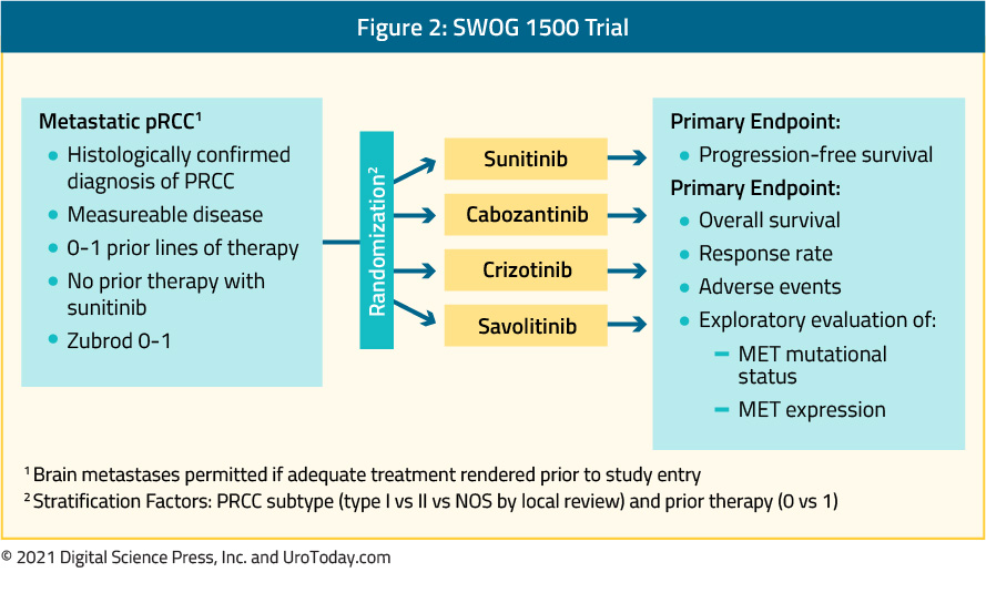
There were 152 patients that were enrolled of whom 5 were ineligible. The included patients had a median age of 66 (range:29-89) and the majority (76%) were male. The vast majority (92%) had not received prior systemic therapy. Median PFS was significantly higher with cabozantinib relative to sunitinib (HR 0.60, 95% CI 0.37-0.97). Objective response rates were also higher with cabozantinib than with sunitinib, crizotinib, and savolitinib, with two complete responses and eight partial responses noted among the 44 patients randomized to cabozantinib. Median OS was 20 months for those receiving cabozantinib and 16.4 months for those receiving sunitinib.
Treatment Selection
As highlighted above, there are a number of treatment approaches which have, in phase III RCTs, demonstrated superiority to sunitinib in first-line treatment of clear cell mRCC including atezolizumab + bevacizumab, nivolumab + ipilimumab, pembrolizumab + axitinib, avelumab + axitinib, nivolumab + cabozantinib, pembrolizumab + lenvatinib. As highlighted in the BIONNIKK trial, a tumor-derived signature may allow for rationale treatment selection, however, prior to this, IMDC risk categories and PD-L1 testing may provide some guidance. Additionally, authors have considered cost-effectiveness analyses to help guide treatment selection29,30. However, as may be expected, varying the assumptions of these models may change the preferred treatment options. Numerous ongoing trials will continue to shape this rapidly evolving disease space and individual treatment choice will depend on the patient, physician, and system factors with guidelines likely to continue to recommend multiple options.
Written by: Zachary Klaassen, MD, MSc, Urologic Oncologist, Assistant Professor Surgery/Urology at the Medical College of Georgia at Augusta University, Georgia Cancer Center
Published Date: March 2021
- Written by: Zachary Klaassen, MD, MSc
- References:
1. Siegel RL, Miller KD, Jemal A. Cancer statistics, 2020. CA: a cancer journal for clinicians. 2020;70(1):7-30.
2. Welch HG, Skinner JS, Schroeck FR, Zhou W, Black WC. Regional Variation of Computed Tomographic Imaging in the United States and the Risk of Nephrectomy. JAMA internal medicine. 2018;178(2):221-227.
3. Motzer RJ, Mazumdar M, Bacik J, Berg W, Amsterdam A, Ferrara J. Survival and prognostic startitifcation of 670 patients with advanced renal cell carcinoma. Journal of Clinical Oncology. 1999;17:2530-2540.
4. Negrier S, Escudier B, Gomez F, et al. Prognostic factors of survival and rapid progression in 782 patients with metastatic renal carcinomas treated by cytokines: a report from the Groupe Francais d'Immunotherapie. Annals of oncology : official journal of the European Society for Medical Oncology / ESMO. 2002;13(9):1460-1468.
5. Motzer RJ, Bacik J, Murphy BA, Russo P, Mazumdar M. Interferon-alfa as a comparative treatment for clinical trials of new therapies against advanced renal cell carcinoma. Journal of clinical oncology : official journal of the American Society of Clinical Oncology. 2002;20(1):289-296.
6. Coppin C, Porzsolt F, Awa A, Kumpf J, Coldman A, Wilt T. Immunotherapy for advanced renal cell cancer. Cochrane Database Syst Rev. 2005(1):CD001425.
7. Dutcher JP, Atkins M, Fisher R, et al. Interleukin-2-based therapy for metastatic renal cell cancer: the Cytokine Working Group experience, 1989-1997. Cancer J Sci Am. 1997;3 Suppl 1:S73-78.
8. Rosenberg SA, Yang JC, White DE, Steinberg SM. Durability of complete responses in patients with metastatic cancer treated with high-dose interleukin-2: identification of the antigens mediating response. Ann Surg. 1998;228(3):307-319.
9. Rini BI, Halabi S, Rosenberg JE, et al. Bevacizumab plus interferon alfa compared with interferon alfa monotherapy in patients with metastatic renal cell carcinoma: CALGB 90206. Journal of clinical oncology : official journal of the American Society of Clinical Oncology. 2008;26(33):5422-5428.
10. Escudier B, Pluzanska A, Koralewski P, et al. Bevacizumab plus interferon alfa-2a for treatment of metastatic renal cell carcinoma: a randomised, double-blind phase III trial. Lancet. 2007;370(9605):2103-2111.
11. Motzer RJ, Hutson TE, Tomczak P, et al. Sunitinib versus interferon alfa in metastatic renal-cell carcinoma. The New England journal of medicine. 2007;356(2):115-124.
12. Motzer RJ, Hutson TE, Cella D, et al. Pazopanib versus sunitinib in metastatic renal-cell carcinoma. The New England journal of medicine. 2013;369(8):722-731.
13. Rini BI, Escudier B, Tomczak P, et al. Comparative effectiveness of axitinib versus sorafenib in advanced renal cell carcinoma (AXIS): a randomised phase 3 trial. Lancet. 2011;378(9807):1931-1939.
14. Hutson TE, Lesovoy V, Al-Shukri S, et al. Axitinib versus sorafenib as first-line therapy in patients with metastatic renal-cell carcinoma: a randomised open-label phase 3 trial. The lancet oncology. 2013;14(13):1287-1294.
15. Choueiri TK, Halabi S, Sanford BL, et al. Cabozantinib Versus Sunitinib As Initial Targeted Therapy for Patients With Metastatic Renal Cell Carcinoma of Poor or Intermediate Risk: The Alliance A031203 CABOSUN Trial. Journal of clinical oncology : official journal of the American Society of Clinical Oncology. 2017;35(6):591-597.
16. Choueiri TK, Hessel C, Halabi S, et al. Cabozantinib versus sunitinib as initial therapy for metastatic renal cell carcinoma of intermediate or poor risk (Alliance A031203 CABOSUN randomised trial): Progression-free survival by independent review and overall survival update. European journal of cancer. 2018;94:115-125.
17. Hudes G, Carducci M, Tomczak P, et al. Temsirolimus, interferon alfa, or both for advanced renal-cell carcinoma. The New England journal of medicine. 2007;356(22):2271-2281.
18. Escudier B, Tannir NM, McDermott D, et al. LBA5 - CheckMate 214: Efficacy and safety of nivolumab 1 ipilimumab (N1I) v sunitinib (S) for treatment-naive advanced or metastatic renal cell carcinoma (mRCC), including IMDC risk and PD-L1 expression subgroups. Annals of Oncology. 2017;28(Supplement 5):621-622.
19. Escudier B, Motzer RJ, Tannir NM, et al. Efficacy of Nivolumab plus Ipilimumab According to Number of IMDC Risk Factors in CheckMate 214. European urology. 2019.
20. McDermott DF, Lee JL, Bjarnason GA, et al. Open-Label, Single-Arm Phase II Study of Pembrolizumab Monotherapy as First-Line Therapy in Patients With Advanced Clear Cell Renal Cell Carcinoma. Journal of clinical oncology : official journal of the American Society of Clinical Oncology. 2021;39(9):1020-1028.
21. Motzer R, Powles T, Atkins M, et al. IMmotion151: A Randomized Phase III Study of Atezolizumab Plus Bevacizumab vs Sunitinib in Untreated Metastatic Renal Cell Carcinoma. Journal of Clinical Oncology. 2018;36(Suppl 6S).
22. Rini BI, Plimack ER, Stus V, et al. Pembrolizumab plus Axitinib versus Sunitinib for Advanced Renal-Cell Carcinoma. The New England journal of medicine. 2019;380(12):1116-1127.
23. Motzer RJ, Penkov K, Haanen J, et al. Avelumab plus Axitinib versus Sunitinib for Advanced Renal-Cell Carcinoma. The New England journal of medicine. 2019;380(12):1103-1115.
24. Motzer R, Alekseev B, Rha SY, et al. Lenvatinib plus Pembrolizumab or Everolimus for Advanced Renal Cell Carcinoma. The New England journal of medicine. 2021.
25. Martinez Chanza N, Xie W, Asim Bilen M, et al. Cabozantinib in advanced non-clear-cell renal cell carcinoma: a multicentre, retrospective, cohort study. The lancet oncology. 2019;20(4):581-590.
26. McDermott DF, Lee JL, Ziobro M, et al. Open-Label, Single-Arm, Phase II Study of Pembrolizumab Monotherapy as First-Line Therapy in Patients With Advanced Non-Clear Cell Renal Cell Carcinoma. Journal of clinical oncology : official journal of the American Society of Clinical Oncology. 2021;39(9):1029-1039.
27. Choueiri TK, Heng DYC, Lee JL, et al. Efficacy of Savolitinib vs Sunitinib in Patients With MET-Driven Papillary Renal Cell Carcinoma: The SAVOIR Phase 3 Randomized Clinical Trial. JAMA Oncol. 2020;6(8):1247-1255.
28. Pal SK, Tangen C, Thompson IM, Jr., et al. A comparison of sunitinib with cabozantinib, crizotinib, and savolitinib for treatment of advanced papillary renal cell carcinoma: a randomised, open-label, phase 2 trial. Lancet. 2021;397(10275):695-703.
29. Su Y, Fu J, Du J, Wu B. First-line treatments for advanced renal-cell carcinoma with immune checkpoint inhibitors: systematic review, network meta-analysis and cost-effectiveness analysis. Ther Adv Med Oncol. 2020;12:1758835920950199.
30. Bensimon AG, Zhong Y, Swami U, et al. Cost-effectiveness of pembrolizumab with axitinib as first-line treatment for advanced renal cell carcinoma. Curr Med Res Opin. 2020;36(9):1507-1517.
First Line Therapy of Metastatic Clear Cell RCC
Background
Kidney cancer represents 5% of all new cancer diagnoses in the United States, with approximately 64,000 new cases and 14,970 deaths in 2018.1,2 The most common type of kidney cancer is renal cell carcinoma (RCC) and the most common histologic subtype of RCC is clear cell RCC, accounting for over 80% of cases.3 RCC is more common in men than women and typically occurs in the sixth to eighth decade of life.1 Localized kidney cancer can often be cured with definitive surgery, with 5-year survival reaching over 90%.4 However, for patients with advanced disease, 5-year survival remains poor at 11.7% and much progress is needed to develop novel therapies for advanced RCC.4Risk Stratification
The current treatment paradigm for metastatic clear cell RCC requires stratification of patients into favorable, intermediate, or poor risk disease. Several validated models for risk stratification exist, including the International Metastatic Renal Cell Carcinoma Database Consortium model (IMDC)5 and Memorial Sloan Kettering Cancer Center model (MSKCC).6 Both criteria include time from diagnosis to systemic treatment of less than one year, performance status, as well as hemoglobin and calcium (Table 1). The main differences between these models are that the MSKCC model includes LDH and the IMDC includes neutrophil and platelet count as unique risk factors. Patients with no risk factors fall into the favorable risk group, patients with one to two risk factors are in the intermediate risk group, and those with three or more risk factors are in the high-risk group for both prognostic models. Contemporary clinical trials have found that drug effectiveness may vary depending on risk stratification which has led the FDA to approve some therapies for only certain risk groups.7 Thus, risk stratification is important for the clinician, not only for discussing prognosis with patients, but also for treatment selection.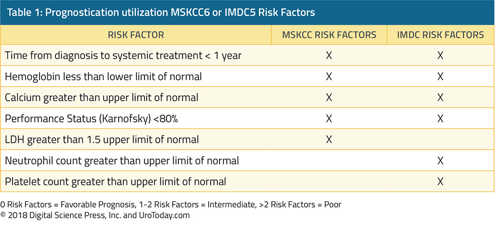
Favorable Risk Patients
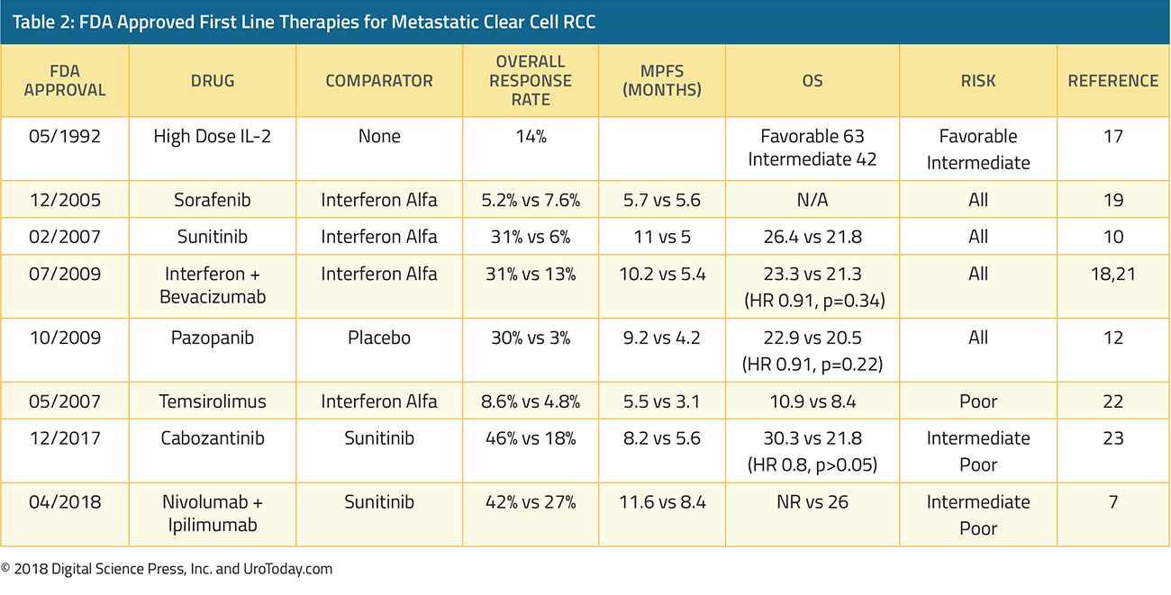
For patients with no adverse risk factors, sunitinib and pazopanib are the preferred first line treatment options, recommended by both the National Comprehensive Cancer Network (NCCN) as well as the European Association of Urology (EAU).8,9 Sunitinib is an oral multikinase inhibitor which targets vascular endothelial growth factor receptors (VEGFR) and platelet-derived growth factor receptors (PDGFR). A phase III trial comparing sunitinib to interferon-α showed that sunitinib significantly improved median progression free survival (11 months vs 5 months) and had a higher overall response rate as well when compared with interferon alfa (31% vs 6%).10 Sunitinib also demonstrated longer overall survival in a follow up study, 26.4 months vs 21.8 months.11 Severe adverse events (grade 3-4 toxicities) were minimal, and the most common adverse events were diarrhea, fatigue, and nausea. Hypertension was one notable side effect of sunitinib, which was not seen with interferon alfa. Based on the data above, sunitinib was granted FDA approval in 2007 and has been the benchmark for many future clinical trials in the mRCC space.Pazopanib is another oral multikinase inhibitor, which targets VEGFR-1,2,3, PDGFR- and , and c-KIT.12 Pazopanib was established as a safe and efficacious therapy for mRCC based on a large randomized, placebo controlled trial in patients who were treatment naive or cytokine pretreated.12 In this trial of 435 patients, pazopanib increased progression-free survival (9.2 months vs 4.2 months) compared with placebo, both in the treatment naïve cohort as well as the cytokine pretreated cohort. The objective response rate to pazopanib was 30% and pazopanib was well tolerated. Unique toxicities of pazopanib included notable grade 3 hepatotoxicity 30% of patients had elevated ALT and 21% had elevated AST. These results granted pazopanib FDA approval in October 2009. A subsequent phase III study (COMPARZ) with 1,110 patients compared sunitinib to pazopanib, and pazopanib was found to be noninferior to sunitinib with respect to progression-free survival and overall survival.13 Median overall survival was 28.4 months in the pazopanib arm compared with 29.3 months in the sunitinib arm. In terms of safety, similar percentages of patients in both sunitinib and pazopanib experienced dose interruptions of one week or greater, 44% and 49% respectively. Patients in the pazopanib arm more frequently discontinued therapy based on abnormal liver function tests. Patients taking sunitinib had a higher risk of abnormal hematologic labs including leukopenia, thrombocytopenia, neutropenia, and anemia.
Additional FDA approved therapies for first line management of favorable risk patients include high dose interleukin 2 (HD IL-2)14-17, interferon plus bevacizumab18, and sorafenib.19 These therapies are less commonly used given their tolerability and toxicity profiles. HD IL-2 deserves special mention here given its ability to induce complete responses in a small subset of patients. A recent abstract describing overall survival from the PROCLAIM database show that of favorable risk patients, median overall survival is 63.3 months and 2 year overall survival is 77.6%.14 Of course, patients who are given HD IL-2 are a very carefully selected robust cohort, and cross-trial comparisons are challenging to make.
Intermediate and Poor Risk
Per the IMDC and MSKCC prognostic models, patients with one or two risk factors are classified as intermediate risk, and poor risk if they have three or more risk factors. Currently, the two major newcomers in this space are cabozantinib and the combination of ipilimumab and nivolumab. Cabozantinib is a multikinase inhibitor of VEGFR, MET, and AXL.20 In the phase II CABOSUN study, 157 intermediate or poor risk patients were randomized to cabozantinib or sunitinib. In this population, cabozantinib increased median progression free survival (8.2 months vs 5.6 months) and improved overall response rate (33% vs to 12%) compared with sunitinib. Both sunitinib and cabozantinib had about a 67% grade 3/4 adverse event rate and had a similar toxicity profile, including fatigue, hypertension, and diarrhea. Sunitinib had a lower incidence of hand foot syndrome and weight loss compared with cabozantinib, but higher rates of neutropenia and thrombocytopenia. Given this data, Cabozantinib obtained FDA approval for the front-line treatment of mRCC in December 2017.The newest therapy to obtain FDA approval is the combination checkpoint inhibitor duo Ipilimumab and Nivolumab (Ipi/Nivo); it was approved in April of 2018, based on the results of CheckMate214.7 CheckMate 214 was a randomized, open-label trial comparing sunitinib with Ipi/Nivo. 1096 patients were enrolled, of which 847 were intermediate or poor risk. This study had a coprimary endpoint of overall survival, objective response rate, and progression free survival among patients who were intermediate or poor risk. The overall response rate was 42% with ipi/nivo vs 27% with sunitinib, with a complete response rate of 9% vs 1%. Median overall survival has not been reached with ipi/nivo vs 26 months for sunitinib. Progression free survival was 11.6 months for ipi/nivo compared with 8.4 months for sunitinib. 46% of patients receiving ipi/nivo experienced grade 3/4 toxicities compared with 63% of patients receiving sunitinib. The most common grade 3/4 adverse events with ipi/nivo was fatigue, diarrhea, and elevated lipase compared with hypertension, hand-foot syndrome, and increased lipase with sunitinib. 35% of patients required high dose corticosteroids for immune related toxicities with ipi/nivo. Â With this data, the European Association of Urology has recommended that ipi/nivo be the new standard of care for patients with intermediate and poor risk disease, and the NCCN has also listed ipi/nivo as a category 1, preferred treatment option for patients with intermediate and poor risk disease.8,9
Future Therapies
Front-line treatment options for mRCC are rapidly evolving.24 Data shown at ASCO and GU ASCO has demonstrated that antiangiogenic agents in combination with checkpoint inhibitors may prolong progression-free survival when compared with kinase inhibitors.25 Several phase III trials exploring this hypothesis are now underway (Table 3). IMmotion 151 is a randomized phase III trial comparing the combination of atezolizumab + bevacizumab vs sunitinib. Progression-free survival was 11.2 months in the intention to treat analysis for patients atezolizumab and bevacizumab, compared with 8.4 months in the sunitinib arm. JAVELIN Renal 100 is investigating avelumab in combination with axitinib and phase I results show a promising overall response rate of 54.5% out of 55 patients.26 KEYNOTE-426 is investigating pembrolizumab in combination with axitinib, compared with sunitinib alone.27 These trials are important and exciting for our patients with mRCC and their future results may alter the standard of care for frontline mRCC.
Published Date: January 29th, 2019
- Written by: Jason Zhu, MD
- References:
- Siegel RL, Miller KD, Jemal A. Cancer statistics, 2018. CA: A Cancer Journal for Clinicians 2018;68:7-30.2018. at https://www.cancer.org/cancer/kidney-cancer/about/key-statistics.html.)
- Moch H, Gasser T, Amin MB, Torhorst J, Sauter G, Mihatsch MJ. Prognostic utility of the recently recommended histologic classification and revised TNM staging system of renal cell carcinoma. Cancer 2000;89:604-14.
- Motzer RJ, Jonasch E, Agarwal N, et al. Kidney cancer, version 2.2017, NCCN clinical practice guidelines in oncology. Journal of the National Comprehensive Cancer Network 2017;15:804-34.
- Heng DY, Xie W, Regan MM, et al. External validation and comparison with other models of the International Metastatic Renal-Cell Carcinoma Database Consortium prognostic model: a population-based study. The lancet oncology 2013;14:141-8.
- Motzer RJ, Mazumdar M, Bacik J, Berg W, Amsterdam A, Ferrara J. Survival and prognostic stratification of 670 patients with advanced renal cell carcinoma. Journal of clinical oncology 1999;17:2530-.
- Motzer RJ, Tannir NM, McDermott DF, et al. Nivolumab plus Ipilimumab versus Sunitinib in Advanced Renal-Cell Carcinoma. New England Journal of Medicine 2018;378:1277-90.
- Motzer R, Jonasch E, Agarwal N. Kidney Cancer: NCCN Evidence Blocks, Version 2.2018, NCCN Clinical Practice Guidelines in Oncology. 2017.
- Powles T, Albiges L, Staehler M, et al. Updated European Association of Urology Guidelines: Recommendations for the Treatment of First-line Metastatic Clear Cell Renal Cancer. European Urology 2018;73:311-5.
- Motzer RJ, Hutson TE, Tomczak P, et al. Sunitinib versus Interferon Alfa in Metastatic Renal-Cell Carcinoma. New England Journal of Medicine 2007;356:115-24.
- Motzer RJ, Hutson TE, Tomczak P, et al. Overall Survival and Updated Results for Sunitinib Compared With Interferon Alfa in Patients With Metastatic Renal Cell Carcinoma. Journal of Clinical Oncology 2009;27:3584-90.
- Sternberg CN, Davis ID, Mardiak J, et al. Pazopanib in Locally Advanced or Metastatic Renal Cell Carcinoma: Results of a Randomized Phase III Trial. Journal of Clinical Oncology 2010;28:1061-8.
- Motzer RJ, Hutson TE, Cella D, et al. Pazopanib versus Sunitinib in Metastatic Renal-Cell Carcinoma. New England Journal of Medicine 2013;369:722-31.
- Fishman MN, Clark JI, Alva AS, et al. Overall survival (OS) by clinical risk category for high dose interleukin-2 (HD IL-2) treated metastatic renal cell cancer (RCC): Data from PROCLAIM. Journal of Clinical Oncology 2018;36:4578-.
- Amin A, White RL. Interleukin-2 in Renal Cell Carcinoma: A Has-Been or a Still-Viable Option? Journal of Kidney Cancer and VHL 2014;1:74-83.
- Alva A, Daniels GA, Wong MKK, et al. Contemporary experience with high-dose interleukin-2 therapy and impact on survival in patients with metastatic melanoma and metastatic renal cell carcinoma. Cancer Immunology, Immunotherapy 2016;65:1533-44.
- Fyfe G, Fisher RI, Rosenberg SA, Sznol M, Parkinson DR, Louie AC. Results of treatment of 255 patients with metastatic renal cell carcinoma who received high-dose recombinant interleukin-2 therapy. Journal of clinical oncology 1995;13:688-96.
- Escudier B, Pluzanska A, Koralewski P, et al. Bevacizumab plus interferon alfa-2a for treatment of metastatic renal cell carcinoma: a randomised, double-blind phase III trial. The Lancet 2007;370:2103-11.
- Escudier B, Szczylik C, Hutson TE, et al. Randomized Phase II Trial of First-Line Treatment With Sorafenib Versus Interferon Alfa-2a in Patients With Metastatic Renal Cell Carcinoma. Journal of Clinical Oncology 2009;27:1280-9.
- Choueiri TK, Escudier B, Powles T, et al. Cabozantinib versus everolimus in advanced renal cell carcinoma. The New England journal of medicine 2015;373:1814-23.
- Escudier B, Bellmunt J, Négrier S, et al. Phase III trial of bevacizumab plus interferon alfa-2a in patients with metastatic renal cell carcinoma (AVOREN): final analysis of overall survival. J Clin Oncol 2010;28:2144-50.
- Hudes G, Carducci M, Tomczak P, et al. Temsirolimus, Interferon Alfa, or Both for Advanced Renal-Cell Carcinoma. New England Journal of Medicine 2007;356:2271-81.
- Choueiri TK, Halabi S, Sanford BL, et al. Cabozantinib Versus Sunitinib As Initial Targeted Therapy for Patients With Metastatic Renal Cell Carcinoma of Poor or Intermediate Risk: The Alliance A031203 CABOSUN Trial. Journal of Clinical Oncology 2017;35:591-7.
- Zarrabi K, Wu S. Current and emerging therapeutic targets for metastatic renal cell carcinoma. Current oncology reports 2018;20:41.
- Motzer RJ, Powles T, Atkins MB, et al. IMmotion151: A Randomized Phase III Study of Atezolizumab Plus Bevacizumab vs Sunitinib in Untreated Metastatic Renal Cell Carcinoma (mRCC). Journal of Clinical Oncology 2018;36:578-.
- Choueiri TK, Larkin JMG, Oya M, et al. First-line avelumab + axitinib therapy in patients (pts) with advanced renal cell carcinoma (aRCC): Results from a phase Ib trial. Journal of Clinical Oncology 2017;35:4504-.
- Rini BI, Powles T, Chen M, Puhlmann M, Atkins MB. Phase 3 KEYNOTE-426 trial: Pembrolizumab (pembro) plus axitinib versus sunitinib alone in treatment-naive advanced/metastatic renal cell carcinoma (mRCC). Journal of Clinical Oncology 2017;35:TPS4597-TPS.
Approach to Adrenal Masses
Brief Overview of Adrenal Physiology
The adrenal is histologically divided into a three zoned cortex and the inner medulla. The adrenal cortex is involved in the multistep process of steroidogenesis. Each region of the cortex (glomerulosa, fasciculata, and reticularis) produces different steroidal end-products (mineralocorticoids, glucocorticoids, and androgens, respectively) as a result of differing ratios and types of enzymes that catalyze steroidogenesis. The adrenal medulla produces catecholamines (norepinephrine, epinephrine, and dopamine) under the control of the sympathetic branch of the autonomic nervous system.Adrenal Pathology
The differential diagnosis of an adrenal mass is broad, including a number of benign and malignant conditions as summarized in Table 1. In patients with bilateral adrenal masses, the differential diagnosis is somewhat shorter but includes metastases, congenital adrenal hyperplasia, adenomas, lymphoma, infectious causes, hemorrhage, pheochromocytoma, and amyloidosis, and ACTH-dependent Cushing's disease.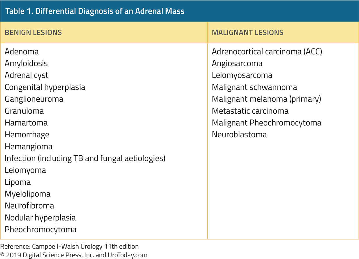
In urologic practice, many adrenal masses represent adrenal incidentalomas, masses >1 cm found on imaging performed for other reasons. While incidentally detected, a relatively large proportion (up to 20%) of these lesions may warrant surgical resection.1 Additionally, more than 10% of these lesions will prove to be biologically active. Therefore, metabolic testing (as detailed below) is recommended for all adrenal incidentalomas.2
Primary adrenal malignancies are uncommon. Adrenocortical carcinoma (ACC) has an incidence of less than 2 per million population.3 While there are associated hereditary syndromes, the majority of ACCs are sporadic. ACC may be biochemically functional or non-functional. Among functional lesions, hypercortisolism is the most common.
From an oncologic perspective, metastases are a much more common cause of adrenal lesions than primary adrenal pathology. Primary cancers with a particular predilection for adrenal metastases including melanoma, lung cancer, renal cell carcinoma, breast cancer, and medullary thyroid cancer.4 However, a wide variety of other cancer may also spread to the adrenal. In patients with known extra-adrenal malignancy, a new adrenal mass is likely to represent metastasis in approximately 50% of cases4. Thus, standard functional assessment is advocated.4
While we will not dwell on it further, a brief mention of congenital adrenal hyperplasia is warranted. This is an autosomal recessive congenital condition characterized by low cortisol production as a result of enzymatic defects in the steroidogenesis pathway. Deficiency in 21-hydroxylase is the cause of nearly 95% of cases. Due to a lack of feedback, there is overproduction of ACTH and resulting overproduction of adrenal androgens. This condition is most often diagnosed and managed in childhood, thus, it will be uncommon as a presentation for adults with newly diagnosed adrenal lesions.
Investigation of Adrenal Lesion
With a newly identified adrenal lesion, there are two primary questions which will guide further management. First, could this mass be malignant? Second, is this mass functional? That is, are there any physical signs and symptoms or biochemical evidence of excess hormonal activity that could be attributed to excess secretion of an adrenally derived hormone.Imaging is warranted (and likely the reason for assessment) for patients with adrenal lesions. Ultrasound is relatively poor at visualizing and characterizing adrenal lesions. Therefore, axial imaging using CT or MRI is advised. Unenhanced CT scan is the first line test of choice. In more than 70% of cases, it is possible to identify adrenal adenomas on the basis of this test alone. Low attenuation (<10 HU) is the characteristic finding on this study. Enhanced CT with adrenal washout protocols may be used where unenhanced CT is unclear. Adenomas exhibit characteristic rapid enhancement washout after administration of CT contrast. MRI is an alternative to CT scan. Again, there are characteristic findings of adrenal adenomas including a loss of signal intensity of out-of-phase sequences.5
Imaging findings help to guide the answer to the question of whether a given adrenal lesion may be malignant. There is a relationship between the size of an adrenal lesion and the likelihood of malignancy. Thus, all lesions larger than 6 cm should be considered malignant until proven otherwise. Due to diagnostic uncertainty, may would advocate resection for lesions 4 cm or larger.1 Additionally, as the incidence of benign lesions increases with age, additional concern should be taken for younger patients with even small adrenal lesions. On axial imaging, ACC exhibit increase attenuation on non-contrast CT, irregular borders and enhancement, and calcification and necrosis.
Functional assessment of adrenal lesions begins with history and physical examination. Cushing's syndrome, caused by excess production of glucocorticoids, may present with central obesity, proximal muscle weakness, thinning of the skin, a so-called buffalo hump, or moon facies. Primary hyperaldosteronism, also known as Conn’s disease, may present with hypertension and hypokalemia. In many patients, hypertension is quite severe with mean blood pressures in the range of 180/1106. Pheochromocytomas, which secrete catecholamines, may present with hypertension, arrhythmia, anxiety, headache, pallor, diaphoresis, and tremor. The classic triad comprised headache, episodic sudden perspiration and tachycardia.7 Adrenocortical carcinoma may produce functional syndromes as described above or may also cause mass-related effects including abdominal fullness, back pain, nausea, and vomiting.
Biochemical assays are employed to confirm functional lesions. For Cushing's syndrome, the diagnosis may be confirmed with a 24-hour urinary free cortisol test or a low-dose dexamethasone suppression test. Following diagnosis, a number of subsequent tests may be performed to ascertain the underlying etiology. While these are typically coordinated by an endocrinologist, they will be briefly summarized here. Determination of serum ACTH (adrenocorticotropic hormone) can distinguish ACTH-dependent Cushing’s from ACTH-independent causes. Among patients with elevated ACTH, determination of the anatomic source, whether pituitary or ectopic, can drive further management. However, modern imaging remains relatively insensitive and non-specific for the detection of both pituitary and ectopic sources of ACTH.8,9 Therefore, direct measurement of venous levels of ACTH in the inferior petrosal sinus following CRH stimulation has been accepted to distinguish pituitary and ectopic sources of ACTH.8 High-dose dexamethasone suppression testing is no longer routinely used.8
Due to the underlying pathophysiology, patients must stop mineralocorticoid receptor antagonist antihypertensives prior to investigation for primary hyperaldosteronism. Further, hypokalemia should be corrected. For these patients, it is critical to determine whether this is a primary process or driven by perturbations in renin levels. Thus, determination of the ratio of serum aldosterone to plasma renin activity (PRA) is critical. This is known as the aldosterone to renin ratio (ARR). For patients with a positive ARR screening test, confirmatory testing typically seeks to identify suppression of aldosterone production following sodium loading. Options include fludrocortisone suppression testing, oral sodium loading, and intravenous saline infusion. Other, less commonly utilized, tests include captopril suppression testing, the furosemide-upright test, and the ACTH stimulation test. However, a number of etiologies may contribute to primary hyperaldosteronism including bilateral or unilateral hyperplasia, adenomas, and tumors. Therefore, following confirmation, subtype investigations may be undertaken among patients who are surgical candidates. This is typically performed with cross-sectional imaging. For patients without identified unilateral nodules, adrenal venous sampling may allow lateralization of the lesion. In the case of a non-diagnostic sampling, other optics including nuclear scintigraphy and postural stimulation testing.
Pheochromocytomas are potentially the most worrisome of functional adrenal lesions given the potential for significant cardiovascular instability if they are not recognized prior to intervention. Evaluation of these masses should include both biochemical and radiographic studies. Biochemical studies assess catecholamines and their metabolites including plasma free metanephrines, catecholamines, urinary fractionated metanephrines, total metanephrines, and vanillylmandelic acid. Each of these tests have varying sensitivity and specificity. Today, most advocate testing of plasma free metanephrine levels10 as this is more sensitive than serum levels of catecholamines. For patients with equivocal findings, use of the clonidine suppression test has been suggested by some.11 Chromogranin A is an alternative confirmatory test though the sensitivity is somewhat poor for this function.
As with all adrenal lesions, imaging of pheochromocytoma begins with computed tomography (CT). Unlike adrenal adenomas, pheochromocytoma typically has an increased attenuation (mean 35 HU).12 Magnetic resonance imaging (MRI) is an alternative. Classically, these lesions have a bright signal, termed the "light bulb" sign. Functional imaging may be undertaken using 18F-FDG PET scanning or metaiodobenzylguanidine (MIBG) scintigraphy.
As hereditary lesions account for nearly 1/3 cases of pheochromocytoma, familial testing has been suggested among patients who have a family history, present at age <50 years, have multiple lesions, malignant pheochromocytoma, or bilateral pheochromocytoma.13
Investigations to assess the functionality of adrenal lesions are summarized in Table 2.

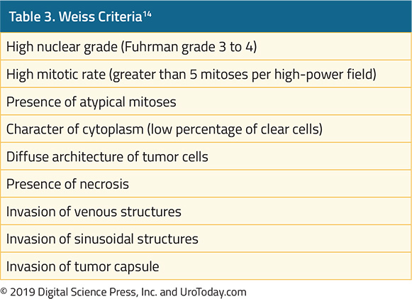
Investigation of suspected ACC should assess excesses of glucocorticoids, sex steroids, catecholamines, and mineralocorticoids. The Weiss pathologic criteria are used to distinguish benign and malignant adrenal lesions (Table 3).14 The presence of three or more of these criteria is highly associated with malignancy.
Treatment of Adrenal Lesions
For patients with small non-functional adrenal lesions with benign imaging findings, surveillance may be appropriate. However, surgery is the mainstay for patients with adrenal lesions. There are particular nuances on the basis of the underlying histology and functional status. In general, laparoscopic adrenalectomy is considered the gold standard as, in experienced hands, oncologic outcomes are equivalent with improved convalescence.For patients with adrenocortical carcinoma, surgical resection is the standard of care. In these cases, wide margins are critical. Thus, for larger tumors with possible adjacent organ involvement, some authors advocate that these cases should be performed open in order to ensure negative margins given the potential need for adjacent organ resection. Unfortunately, recurrence is common following even aggressive resection. Radiotherapy can be used in an adjuvant setting for patients with positive margins and for treatment of bone or central nervous system metastases. Systemic therapy may be undertaken with mitotane, a synthetic derivative of DDT.
For patients with Cushing's disease, the management varies widely based on underlying etiology. The overall goals included correction of the cortisol excess, restoration of the underlying hormonal axis, and management of the sequelae. Approaches to this, depending on underlying etiology, include weaning of exogenous steroids, transsphenoidal resection of pituitary lesions, unilateral or bilateral adrenalectomy, resection of ectopic sources of ACTH, and medical therapy with blockers of steroidogenesis.
Treatment of primary aldosteronism seeks to control blood pressure and prevent sequelae of hormonal excess. This may be accomplished medically or surgically depending on the underlying cause and patient suitability for operation. Medical treatment may be undertaken with aldosterone receptor antagonists such as spironolactone or eplerenone.
Pheochromocytoma is primarily a surgical disease. However, extensive medical consultation and optimization is required to prevent significant intraoperative cardiovascular complications. Further, these patients are at risk of cardiomyopathy and, therefore, consultation with a cardiologist or anesthesiologist prior to surgery is advisable. Catecholamine blockade is required prior to surgery on pheochromocytoma. Classically, this has been achieved with the non-competitive alpha-blocker phenoxybenzamine. However, selective reversible alpha-blockers including doxazosin or terazosin are alternatives. Following alpha-blockade, beta-blockade may be undertaken due to the risk of reflex tachycardia or arrhythmia.13 An alternative to alpha- and beta-blockade which has been proposed utilized calcium channel blockade.15 Finally, catecholamine synthesis blockade through the use of alpha-methyltyrosine (metyrosine) may be added. In the perioperative period, repletion of the intravascular volume is critical. This may be achieved through liberal consumption of salt and liquid or intravenous resuscitation. Careful postoperative monitoring is key as these patients are at risk for hypotension and hypoglycemia. Additionally, as these lesions have a predilection for recurrence, ongoing monitoring is required.
Published Date: January 28th, 2019
- References:
- Young WF, Jr. Clinical practice. The incidentally discovered adrenal mass. The New England journal of medicine 2007;356:601-10.
- Grumbach MM, Biller BM, Braunstein GD, et al. Management of the clinically inapparent adrenal mass ("incidentaloma"). Ann Intern Med 2003;138:424-9.
- Fassnacht M, Kroiss M, Allolio B. Update in adrenocortical carcinoma. The Journal of clinical endocrinology and metabolism 2013;98:4551-64.
- Lenert JT, Barnett CC, Jr., Kudelka AP, et al. Evaluation and surgical resection of adrenal masses in patients with a history of extra-adrenal malignancy. Surgery 2001;130:1060-7.
- Namimoto T, Yamashita Y, Mitsuzaki K, et al. Adrenal masses: quantification of fat content with double-echo chemical shift in-phase and opposed-phase FLASH MR images for differentiation of adrenal adenomas. Radiology 2001;218:642-6.
- Young WF, Jr., Klee GG. Primary aldosteronism. Diagnostic evaluation. Endocrinol Metab Clin North Am 1988;17:367-95.
- Bravo EL, Tagle R. Pheochromocytoma: state-of-the-art and future prospects. Endocr Rev 2003;24:539-53.
- Porterfield JR, Thompson GB, Young WF, Jr., et al. Surgery for Cushing's syndrome: an historical review and recent ten-year experience. World J Surg 2008;32:659-77.
- Newell-Price J, Bertagna X, Grossman AB, Nieman LK. Cushing's syndrome. Lancet 2006;367:1605-17.
- Lenders JW, Pacak K, Walther MM, et al. Biochemical diagnosis of pheochromocytoma: which test is best? JAMA : the Journal of the American Medical Association 2002;287:1427-34.
- Eisenhofer G, Goldstein DS, Walther MM, et al. Biochemical diagnosis of pheochromocytoma: how to distinguish true- from false-positive test results. The Journal of clinical endocrinology and metabolism 2003;88:2656-66.
- Motta-Ramirez GA, Remer EM, Herts BR, Gill IS, Hamrahian AH. Comparison of CT findings in symptomatic and incidentally discovered pheochromocytomas. AJR Am J Roentgenol 2005;185:684-8.
- Pacak K. Preoperative management of the pheochromocytoma patient. The Journal of Clinical Endocrinology and metabolism 2007;92:4069-79.
- Weiss LM. Comparative histologic study of 43 metastasizing and nonmetastasizing adrenocortical tumors. Am J Surg Pathol 1984;8:163-9.
- Ulchaker JC, Goldfarb DA, Bravo EL, Novick AC. Successful outcomes in pheochromocytoma surgery in the modern era. The Journal of Urology 1999;161:764-7.
Systemic Therapy for Advanced Renal Cell Carcinoma
As emphasized in the article on Malignant Renal Tumors, clear cell renal cell carcinoma (ccRCC) is the most common histologic subtype of renal cell carcinoma (RCC). Likely due to its much higher prevalence, the vast majority of systemic therapies for RCC have been investigated among patients with ccRCC. Historically, treatment for metastatic RCC (mRCC) had been limited to cytokine therapies (interleukin-2 and interferon-alfa). However, the development of tyrosine kinase inhibitors (TKIs), which target vascular endothelial growth factors (VEGF), and mammalian target of rapamycin (mTOR) inhibitors have replaced cytokine-based therapies as the standard of care. More recently, immunotherapy-based approaches using checkpoint inhibitors have demonstrated significant benefits and have joined the repertoire of available agents for patients with metastatic RCC.
Cytokine Therapies for Advanced ccRCC
The host immune system has long been implicated with RCC tumor biology. As a result, modulators of the immune system were among the first therapeutic targets for advanced ccRCC.Interferon-α was one of the first cytokines assessed for the treatment of metastatic ccRCC. Interferons have a range of biologic functions, including immunomodulation. Early data demonstrated response rates in the range of 10 to 15% for patients treated with interferon-α.4 Compared with other available systemic therapies available at the time, interferon therapy conferred a survival benefit.5
An alternative form of immunologic modulation was examined using interleukin-2. While response rates were similar to interferon-based therapies (~15 to 20%)6, interleukin-2 was distinct in that durable complete responses were observed in approximately 7 to 9% of patients.7 On the basis of these data, high-dose IL-2 was approved by the U.S. Food and Drug Administration (FDA) in 1992. However, IL-2 is associated with considerable toxicity which has limited its widespread utilization. Most worrisome is vascular leak syndrome which leads to intravascular depletion, hypovolemia, respiratory compromise and multi-organ failure. Alternatives to the high-dose intravenous bolus administration were explored but lead to worse oncologic outcomes. Thus, high-dose IL-2 is the only recommended approach for patients undergoing cytokine therapy.
Subsequently, combinations of interferon and interleukin therapies were explored. These demonstrated some improvement in response rate but no difference in overall survival.8 Combination therapy resulted in significantly increased toxicity compared to monotherapy with either agent.
With the introduction of VEGF and mTOR targeting agents, interferon is no longer utilized as first-line therapy. However, IL-2 remains an available, though not widely utilized, option on the basis of its ability to induce durable complete responses which these new agents lack.
Inhibitors of the VEGF Pathway for Advanced ccRCC
Based on work into the molecular biology underlying ccRCC led to “rational targeted therapeutics” including targeting of the VEGF pathway.The first inhibitor of the VEGF pathway used in clinical trials was bevacizumab, a humanized monoclonal antibody against VEGF-A. While this approach was first explored in patients who had progressed on cytokine-based therapies, it was soon evaluated head-to-head against interferon in previously untreated patients.9,10 The addition of bevacizumab to interferon resulted in significant improvements in response rate and progression-free survival. Today, bevacizumab is uncommonly used as monotherapy in untreated patients but is considered as second-line therapy in patients who have failed prior therapy with tyrosine kinase inhibitors.
Tyrosine-kinase inhibitors also target the VEGF pathway, through inhibition of a combination of VEGFR-2, PDGFR-β, raf-1 c-Kit, and Flt3 (sunitinib and sorafenib). In 2006, sorafenib was shown to have biologic activity in ccRCC. Subsequent studies demonstrated improvements in progression-free survival compared with placebo in patients who have previously failed cytokine therapy and improvements in tumor regression compared to interferon in previously untreated patients. Despite FDA approval, sorafenib is rarely used as first-line therapy today. More widely used is sunitinib. As with agents discussed, sunitinib was first evaluated among patients who had previously received cytokine treatment. Subsequently, it was compared to interferon-α in a large phase III randomized trial.11 While the initial analysis demonstrated significant improvement in progression-free survival and overall response rate, subsequent follow-up has demonstrated a strong trend towards improved overall survival. On account of these data, sunitinib is widely used as first-line treatment of RCC.
Tyrosine-kinase inhibitors exhibit a class-based toxicity profile including gastrointestinal events, dermatologic complications including hand-foot desquamation, hypertension, and general malaise. However, quality of life appears to be better with these agents than with interferon.11
Subsequently, a number of more targeted tyrosine kinase inhibitors have been developed with the goal to decrease the toxicity of this treatment strategy. Such agents include pazopanib, axitinib, and tivozanib. Comparative data of pazopanib and sunitinib have demonstrated non-inferior oncologic outcomes with decreased toxicity among patients receiving pazopanib.12 Axitinib was evaluated as second-line therapy compared to sorafenib among patients who had previously received sunitinib, bevacizumab, temsirolimus, or cytokine therapy. Axitinib was associated with improved progression-free survival; on the basis of these data, this agent was approved for second-line therapy of metastatic RCC.13 Finally, tivozanib has been compared to sorafenib among patients who had not previously received VEGF or mTOR-targeting therapies. While this study demonstrated tivozanib’s activity, it was not FDA approved and is therefore not used.
Most recently, a multikinase inhibitor, cabozantinib, has been approved for the first-line treatment of mRCC. In the phase II CABOSUN trial, cabozantinib demonstrated improved progression-free survival compared to sunitinib.14 However, these results have proven controversial, with a number of concerns raised including a potential exaggerated effect due to the poor efficacy of sunitinib compared to what would be expected based on previous reports.15
Despite the efficacy of VEGF targeted therapies, resistance to VEGF-inhibition almost inevitably results. Therefore, research into the development of these resistance mechanisms and ways to target these pathways has been undertaken. Agents including nintedanib and dovitinib have been explored though these are not yet in routine practice.
Inhibitors of mTOR for Advanced ccRCC
Mammalian target of rapamycin (mTOR) plays a key role in regulating HIF-α, thus modulating the pathway between abnormalities in VHF and proliferation. Two analogous of sirolimus have demonstrated efficacy in treating advanced RCC, temsirolimus and everolimus.A three-arm trial comparing temsirolimus, interferon, and the combination was undertaken among patients with pre-defined poor risk disease who had not previously received systemic therapy for RCC.16 This demonstrated improvements in progression-free survival and overall survival for patients receiving temsirolimus. Notably, the combination arm did not offer a benefit compared to interferon alone. Unlike temsirolimus which must be administered intravenously, everolimus is an oral agent. Among patients progressing on sunitinib and/or sorafenib, everolimus demonstrated significantly improved progression-free survival compared to placebo.17
Checkpoint Inhibitors for Advanced ccRCC
The immunologic basis for treatment of advanced RCC has been well established, including the aforementioned cytokine therapies. Recently, immune checkpoint inhibitors have been examined in the treatment of advanced RCC. Two particular regimes warrant focus – nivolumab and ipilimumab and atezolizumab.First presented at ESMO in the fall of 2017 and subsequently published in the spring of 2018, CheckMate 214 demonstrated an overall survival (OS) benefit for first-line nivolumab plus ipilimumab vs. sunitinib.18 More details regarding this study may be found in the UroToday coverage of ESMO 2017. In short, among the subgroup of patients with intermediate or poor-risk RCC, treatment with nivolumab plus ipilimumab resulted in significantly improved overall response rate, comparable progression-free survival, and significantly improved overall survival.
Similarly, first presented at GU ASCO in the spring of 2018 and subsequently published, IMmotion151 reported a progression-free survival (PFS) benefit for first-line atezolizumab + bevacizumab vs. sunitinib.19 This regime was active with a significant benefit in progression-free survival among the whole cohort of patients, as well as a subset of PD-L1+ patients. More details regarding this study may be found in the UroToday coverage of GU ASCO 2018.
These trials are notable in that they demonstrated improved outcomes in first-line treatment, compared to the current standard of care, sunitinib.
Other Agents for Advanced ccRCC
Numerous chemotherapeutic agents have been explored in ccRCC. These include 5-FU, gemcitabine, vinblastine, bleomysin, and platinums. Meta-analyses of these data demonstrate poor response20 and thus cytotoxic chemotherapy is not indicated in the treatment of advanced RCC. Similarly, hormonal therapies including medroxyprogesterone have been explored but have no role in modern management of advanced RCC.Treatment of Advanced non-ccRCC
There is a relative dearth of data for treatment of advanced non-clear cell RCC. Therefore, patients with these tumors may receive agents on the basis of their activity in ccRCC. However, VEGF-receptor inhibitors have been shown to have relatively low activity in patients with papillary RCC.21 Responses were somewhat better in patients with chromophobe RCC. Temsirolimus and everolimus appear to have some activity in patients with non-clear cell histology. Similarly, nivolumab monotherapy appears to have biologic activity in patients with non-ccRCC.22Integration of treatment options for patients with advanced RCC
With so many active agents available for the treatment of advanced RCC, it may be difficult to ascertain which treatment to offer patients who present in clinic. There are a number of ways to approach this issue – first, one may take a quantitative approach, utilizing the available comparative data in a network meta-analysis; second, one may rely upon eminence, as in expert-informed guidelines; finally, one may rely on individual clinical experience. In this setting, all three options are available.First, assessing this in a quantitative fashion, we performed a network meta-analysis of agents for the treatment of advanced RCC.23 While there are limitations to this approach including the reliance on the assumption of transitivity between studies, interesting conclusions may be drawn. First, assessing progression-free survival, we found that it was highly likely (91% chance) that cabozantinib provided the greatest benefit. However, when assessing overall survival, nivolumab plus ipilimumab had the highest likelihood of being the preferred treatment choice. Finally, when assessing adverse events, it was highly likely that nivolumab plus ipilimumab had the most favorable toxicity profile.
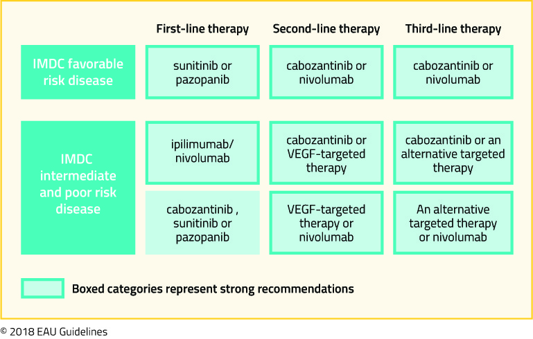
Second, considering a panel of expert opinion, the European Association of Urology updated their guidelines on the treatment of renal cell carcinoma recently. Their recommendations are highlighted in the following figure, taken from the EAU guidelines:
Finally, we may rely on the guidance of individual clinical experience. Anil Kapoor, MD who has extensive experience in the treatment of both localized and advanced RCC, offered his treatment approach recently to UroToday.
Published Date: January 21st, 2019
- References:
- Siegel RL, Miller KD, Jemal A. Cancer statistics, 2018. CA: a cancer journal for clinicians. 2018;68(1):7-30.
- Motzer RJ, Mazumdar M, Bacik J, Berg W, Amsterdam A, Ferrara J. Survival and prognostic stratification of 670 patients with advanced renal cell carcinoma. Journal of Clinical Oncology. 1999;17:2530-2540.
- Negrier S, Escudier B, Gomez F, et al. Prognostic factors of survival and rapid progression in 782 patients with metastatic renal carcinomas treated by cytokines: a report from the Groupe Francais d'Immunotherapie. Annals of oncology : official journal of the European Society for Medical Oncology / ESMO. 2002;13(9):1460-1468.
- Motzer RJ, Bacik J, Murphy BA, Russo P, Mazumdar M. Interferon-alfa as a comparative treatment for clinical trials of new therapies against advanced renal cell carcinoma. Journal of clinical oncology : official journal of the American Society of Clinical Oncology. 2002;20(1):289-296.
- Coppin C, Porzsolt F, Awa A, Kumpf J, Coldman A, Wilt T. Immunotherapy for advanced renal cell cancer. Cochrane Database Syst Rev. 2005(1):CD001425.
- Dutcher JP, Atkins M, Fisher R, et al. Interleukin-2-based therapy for metastatic renal cell cancer: the Cytokine Working Group experience, 1989-1997. Cancer J Sci Am. 1997;3 Suppl 1:S73-78.
- Rosenberg SA, Yang JC, White DE, Steinberg SM. Durability of complete responses in patients with metastatic cancer treated with high-dose interleukin-2: identification of the antigens mediating response. Ann Surg. 1998;228(3):307-319.
- Negrier S, Escudier B, Lasset C, et al. Recombinant human interleukin-2, recombinant human interferon alfa-2a, or both in metastatic renal-cell carcinoma. Groupe Francais d'Immunotherapie. The New England journal of medicine. 1998;338(18):1272-1278.
- Rini BI, Halabi S, Rosenberg JE, et al. Bevacizumab plus interferon alfa compared with interferon alfa monotherapy in patients with metastatic renal cell carcinoma: CALGB 90206. Journal of clinical oncology: official journal of the American Society of Clinical Oncology. 2008;26(33):5422-5428.
- Escudier B, Pluzanska A, Koralewski P, et al. Bevacizumab plus interferon alfa-2a for treatment of metastatic renal cell carcinoma: a randomised, double-blind phase III trial. Lancet. 2007;370(9605):2103-2111.
- Motzer RJ, Hutson TE, Tomczak P, et al. Sunitinib versus interferon alfa in metastatic renal-cell carcinoma. The New England journal of medicine. 2007;356(2):115-124.
- Motzer RJ, Hutson TE, Cella D, et al. Pazopanib versus sunitinib in metastatic renal-cell carcinoma. The New England journal of medicine. 2013;369(8):722-731
- Rini BI, Escudier B, Tomczak P, et al. Comparative effectiveness of axitinib versus sorafenib in advanced renal cell carcinoma (AXIS): a randomised phase 3 trial. Lancet. 2011;378(9807):1931-1939.
- Choueiri TK, Halabi S, Sanford BL, et al. Cabozantinib Versus Sunitinib As Initial Targeted Therapy for Patients With Metastatic Renal Cell Carcinoma of Poor or Intermediate Risk: The Alliance A031203 CABOSUN Trial. Journal of clinical oncology: official journal of the American Society of Clinical Oncology. 2017;35(6):591-597
- Buti S, Bersanelli M. Is Cabozantinib Really Better Than Sunitinib As First-Line Treatment of Metastatic Renal Cell Carcinoma? Journal of clinical oncology: official journal of the American Society of Clinical Oncology. 2017;35(16):1858-1859.
- Hudes G, Carducci M, Tomczak P, et al. Temsirolimus, interferon alfa, or both for advanced renal-cell carcinoma. The New England journal of medicine. 2007;356(22):2271-2281.
- Motzer RJ, Escudier B, Oudard S, et al. Efficacy of everolimus in advanced renal cell carcinoma: a double-blind, randomised, placebo-controlled phase III trial. Lancet. 2008;372(9637):449-456.
- Escudier B, Tannir NM, McDermott D, et al. LBA5 - CheckMate 214: Efficacy and safety of nivolumab 1 ipilimumab (N1I) v sunitinib (S) for treatment-naive advanced or metastatic renal cell carcinoma (mRCC), including IMDC risk and PD-L1 expression subgroups. Annals of Oncology. 2017;28(Supplement 5):621-622.
- Motzer R, Powles T, Atkins M, et al. IMmotion151: A Randomized Phase III Study of Atezolizumab Plus Bevacizumab vs Sunitinib in Untreated Metastatic Renal Cell Carcinoma. Journal of Clinical Oncology. 2018;36(Suppl 6S).
- Yagoda A, Abi-Rached B, Petrylak D. Chemotherapy for advanced renal-cell carcinoma: 1983-1993. Semin Oncol. 1995;22(1):42-60.
- Choueiri TK, Plantade A, Elson P, et al. Efficacy of sunitinib and sorafenib in metastatic papillary and chromophobe renal cell carcinoma. Journal of clinical oncology : official journal of the American Society of Clinical Oncology. 2008;26(1):127-131
- Koshkin VS, Barata PC, Zhang T, et al. Clinical activity of nivolumab in patients with non-clear cell renal cell carcinoma. J Immunother Cancer. 2018;6(1):9.
- Wallis CJD, Klaassen Z, Bhindi B, et al. First-line Systemic Therapy for Metastatic Renal Cell Carcinoma: A Systematic Review and Network Meta-analysis. European urology. 2018;74(3):309-321.
Treatment of Metastatic Non-Clear Cell RCC
Background
Kidney cancer is the 12th most common cancer in the world, with over 300,000 new cases annually, of which 65,340 new cases will be diagnosed in the United States in 2018.1 The incidence of renal cell carcinoma (RCC) varies substantially based on the country – rates of RCC are higher in Europe and North America and much lower in Asia and South America.2
Most kidney cancers (>90%) are renal cell carcinomas, and of renal cell carcinomas, the majority of cases (80%) will be the clear cell subtype.3 Of the remaining 20% of cases, the two major histological subtypes are papillary (10-14%) and chromophobe (5%)3,4, but also include collecting duct, translocation carcinoma, medullary carcinoma, and unclassified RCC. These histological subtypes are distinct from clear cell carcinoma and independently predict for survival.3 For example, after controlling for TNM status, age, gender, and tumor size, patients with early stage clear cell RCC are more than twice as likely to die of RCC than patients with papillary or chromophobe RCC.3 Some of these subtypes also have unique risk factors. For example, renal medullary carcinomas are an aggressive non-clear cell carcinoma that are almost exclusively associated with patients with sickle cell trait.5
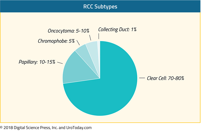
Non-clear cell RCCs (ncRCC) also have unique mutational landscapes.6 For example, MET mutations can be found in 15-30% of papillary RCCs.6,7 Papillary RCCs also have a higher mutation rate compared with chromophobe RCCs and renal oncocytomas. Durinck et al evaluated 167 primary human tumors including papillary, chromophobe and translocation subtypes and were able to use a five gene set to help molecularly distinguish between chromophobe carcinoma, renal oncocytoma and papillary carcinoma.6
Treatment
Given the rarity of non-clear cell RCC (ncRCC), there is a paucity of large randomized phase III trials to help guide the optimal therapy for ncRCC. Some trials for ncRCC have had to stop due to slow accrual. Given this lack of data, patients are encouraged to participate in clinical trials when they are available and appropriate. Below is a summary of the most common systemic therapies in use for ncRCC.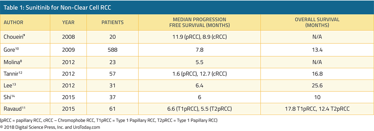
Sunitinib
Single agent sunitinib has been evaluated as part of an expanded access trial as well as several small phase II trials (Table 1). In a single arm phase II clinical trial, 23 patients were given sunitinib 50 mg in cycles of four weeks on followed by 2 weeks off. The trial was stopped early due to slow accrual and the median progression free survival (PFS) was 5.5 months.8 In a another phase II trial evaluating both sunitinib and sorafenib, 19 patients with ncRCC were given sunitinib – median PFS was 11.9 months in the papillary arm and 8.9 months in the chromophobe arm.9 The largest of these studies was reported by Gore et al, which included 588 ncRCC patients who received sunitinib as part of the expanded access study encompassing 4564 RCC patients who received sunitinib.10 In their study, 11% of patients had an objective response and median PFS was 7.8 months. Some trials have stratified outcomes by histological subtypes such as chromophobe or papillary, and one trial reported results broken down by type 1 papillary RCC vs type 2 papillary RCC11. Overall, most studies demonstrated that sunitinib led to a median PFS of about 6-7 months for ncRCC.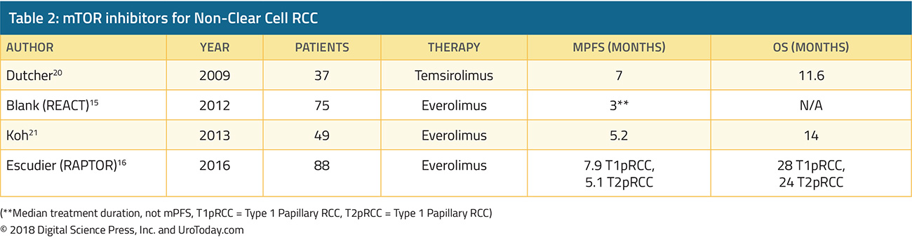
Everolimus and Temsirolimus
mTOR inhibitors Everolimus and Temsirolimus have been evaluated by a few phase II trials for ncRCC (Table 2). Everolimus was evaluated in a subgroup of REACT (RAD001 Expanded Access Clinical Trial in RCC) and in RAPTOR (RAD001 in Advanced Papillary Tumor Program in Europe).15,16 In REACT, 1.3% of patients with ncRCC had a partial response and 49.3% had stable disease.15 In RAPTOR, the median progression free survival (mPFS) was 4.1 months and median OS was 21.4 months. For patients with chromophobe RCC, mTOR directed therapy may be especially effective – case reports show partial responses and stable disease to both Everolimus or Temsirolimus.17,18 This may be due to the hypothesis that a high number of chromophobe RCC’s have PI3K-mTOR pathway activation as well as frequent TSC1/TSC2 mutations which may sensitize these tumors to mTOR inhibition.19
Sunitinib vs Everolimus
Sunitinib and everolimus have also been examined head to head. The largest trial was ASPEN (Everolimus versus sunitinib for patients with metastatic non-clear-cell renal cell carcinoma), a multicenter open-label, randomized phase II trial which randomized 108 patients to receive either sunitinib or everolimus.22 The primary endpoint of this study was progression free survival. The majority of patients had papillary histology (65%). Median overall survival was 13.2 months in the everolimus group and 31.5 months in the sunitinib group. However, overall survival was not statistically different between the two treatment groups (HR 1.12, 95%CI 0.7-2.1, p=0.6). A second study, ESPN (Everolimus Versus Sunitinib Prospective Evaluation in Metastatic Non–Clear Cell Renal Cell Carcinoma) came to a similar conclusion and found that the median overall survival was 16.2 months with sunitinib and 14.9 months with everolimus (p=0.18).23
While overall survival between everolimus and sunitinib were not statistically different for the unselected cohorts, ASPEN did find differences in objective responses between the different subtypes, suggesting that each subtype of ncRCC may respond differently to therapies. In ASPEN, 24% (8/33) of papillary RCCs achieved a partial or complete radiographic response on sunitinib compared with 5% of patients on everolimus (2/37). Interestingly, clinical outcomes after receipt of either sunitinib or everolimus also varied based on risk stratification. Patients with good or intermediate risk had improved median progression free survival (mPFS) with first line sunitinib than everolimus. However, patients with poor risk had improved PFS with everolimus over sunitinib. This is concurrent with the ARCC trial, which also demonstrated improved overall survival with an mTOR inhibitor (temsirolimus) in poor risk patients with ncRCC.24
A meta-analysis of five studies (ESPN, ASPEN, RECORD3, ARCC, and SWOG1107) found a nonstatistical trend favoring sunitinib over everolimus for ncRCC but does note that there is considerable patient heterogeneity in these small studies and there was no statistical difference in PFS between these two therapies.25 In the absence of clinical trial options, sunitinib is a reasonable first line choice for treatment naïve patients with ncRCC, especially for those with papillary RCC or MSKCC good risk RCC.
Special Populations
It is well recognized that ncRCC is a heterogenous mix of patients which respond differently to therapies. Thus, there have been a few biomarker or histology driven trials looking at specific subsets of ncRCC. For example, for patients with hereditary leiomyomatosis and papillary RCC, a phase II study of 41 patients found that patients treated bevacizumab plus erlotinib had a median PFS of 24.2 months, compared to 7.4 months for patients with sporadic papillary RCC.26 Patients with papillary RCC frequently have MET mutations and a variety of MET inhibitors including crizotinib, savolitinib, and cabozantinib are being evaluated in clinical trials.27-29 For example, in a study of 41 patients with type 1 papillary RCC, of the 4 patients with MET+ tumors, 2 had achieved a partial response and one had stable disease with crizotinib.28 In a study with Savolitinib, another selective MET inhibitor, patients with MET “driven” tumors had a median PFS of 6.2 months, compared with 1.7 months for MET independent tumors.29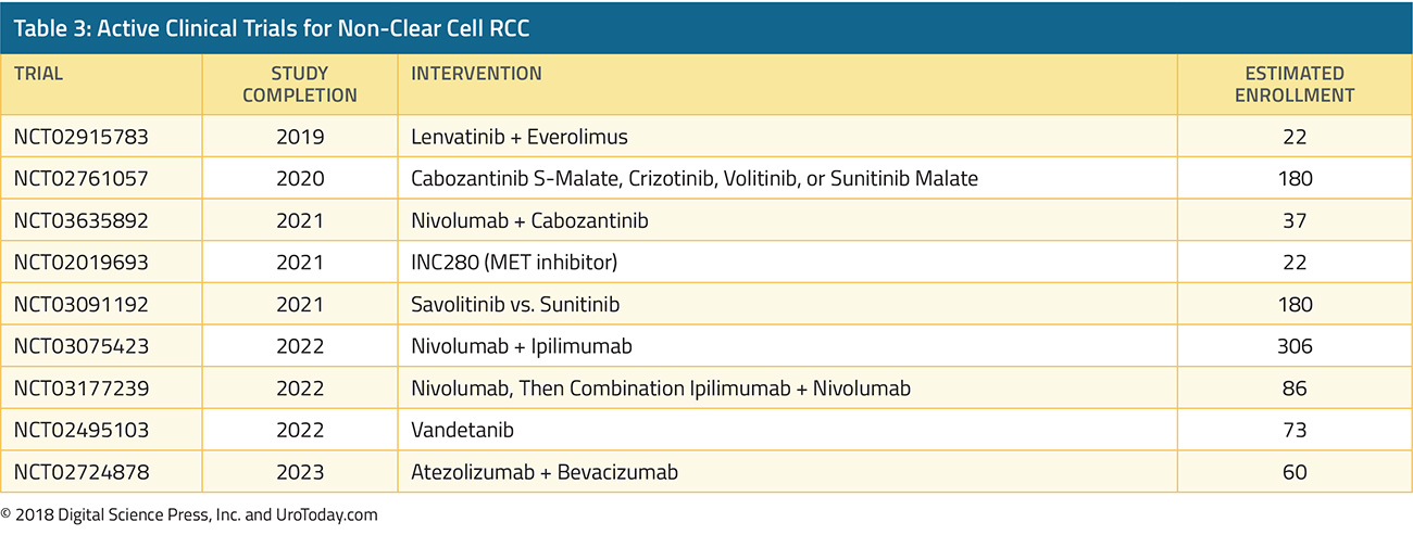
Future Direction
A number of active clinical trials are in progress, investigating various MET inhibitors as well as checkpoint inhibitors for ncRCC (Table 3). Preliminary data suggest that PD-1 or PD-L1 blockade may have some activity in this population.30,31 Given the dearth of data and rarity of ncRCC, it is important to consider these patients for clinical trials, whenever possible.
Published Date: November 29th, 2018
- Written by: Jason Zhu, MD
- References:
1. Motzer R, Jonasch E, Agarwal N. Kidney Cancer: NCCN Evidence Blocks, Version 2.2018, NCCN Clinical Practice Guidelines in Oncology. 2017.
2. Chow W-H, Dong LM, Devesa SS. Epidemiology and risk factors for kidney cancer. Nature Reviews Urology 2010;7:245.
3. Leibovich BC, Lohse CM, Crispen PL, et al. Histological Subtype is an Independent Predictor of Outcome for Patients With Renal Cell Carcinoma. The Journal of Urology 2010;183:1309-16.
4. Moch H, Gasser T, Amin MB, Torhorst J, Sauter G, Mihatsch MJ. Prognostic utility of the recently recommended histologic classification and revised TNM staging system of renal cell carcinoma. Cancer 2000;89:604-14.
5. Shetty A, Matrana MR. Renal Medullary Carcinoma: A Case Report and Brief Review of the Literature. The Ochsner Journal 2014;14:270-5.
7. Carlo MI, Khan N, Chen Y, et al. The genomic landscape of metastatic non-clear cell renal cell carcinoma. American Society of Clinical Oncology; 2017.
8. Molina AM, Feldman DR, Ginsberg MS, et al. Phase II trial of sunitinib in patients with metastatic non-clear cell renal cell carcinoma. Investigational New Drugs 2012;30:335-40.
9. Choueiri TK, Plantade A, Elson P, et al. Efficacy of Sunitinib and Sorafenib in Metastatic Papillary and Chromophobe Renal Cell Carcinoma. Journal of Clinical Oncology 2008;26:127-31.
10. Gore ME, Szczylik C, Porta C, et al. Safety and efficacy of sunitinib for metastatic renal-cell carcinoma: an expanded-access trial. The Lancet Oncology 2009;10:757-63.12. Tannir NM, Plimack E, Ng C, et al. A phase 2 trial of sunitinib in patients with advanced non–clear cell renal cell carcinoma. European urology 2012;62:1013-9.
13. Lee JL, Ahn JH, Lim HY, et al. Multicenter phase II study of sunitinib in patients with non-clear cell renal cell carcinoma. Annals of Oncology 2012;23:2108-14.
14. Shi H-Z, Tian J, Li C-L. Safety and efficacy of sunitinib for advanced non-clear cell renal cell carcinoma. Asia-Pacific Journal of Clinical Oncology 2015;11:328-33.
15. Blank CU, Bono P, Larkin JMG, et al. Safety and efficacy of everolimus in patients with non-clear cell renal cell carcinoma refractory to VEGF-targeted therapy: Subgroup analysis of REACT. Journal of Clinical Oncology 2012;30:402-.
17. Larkin JMG, Fisher RA, Pickering LM, et al. Chromophobe Renal Cell Carcinoma With Prolonged Response to Sequential Sunitinib and Everolimus. Journal of Clinical Oncology 2011;29:e241-e2.
18. Shuch B, Vourganti S, Friend JC, Zehngebot LM, Linehan WM, Srinivasan R. Targeting the mTOR pathway in chromophobe kidney cancer. Journal of Cancer 2012;3:152.
19. Maroto P, Anguera G, Roldan-Romero JM, et al. Biallelic TSC2 Mutations in a Patient With Chromophobe Renal Cell Carcinoma Showing Extraordinary Response to Temsirolimus. Journal of the National Comprehensive Cancer Network 2018;16:352-8.
20. Dutcher JP, Atkins M, Fisher R, et al. Interleukin-2-based therapy for metastatic renal cell cancer: the Cytokine Working Group experience, 1989-1997. The cancer journal from Scientific American 1997;3:S73-8.
21. Koh Y, Lim HY, Ahn JH, et al. Phase II trial of everolimus for the treatment of nonclear-cell renal cell carcinoma. Annals of Oncology 2013;24:1026-31.
24. Hudes G, Carducci M, Tomczak P, et al. Temsirolimus, Interferon Alfa, or Both for Advanced Renal-Cell Carcinoma. New England Journal of Medicine 2007;356:2271-81.
26. Srinivasan R, Su D, Stamatakis L, et al. 5 Mechanism based targeted therapy for hereditary leiomyomatosis and renal cell cancer (HLRCC) and sporadic papillary renal cell carcinoma: interim results from a phase 2 study of bevacizumab and erlotinib. European Journal of Cancer 2014;50:8.
27. Martinez Chanza N, Bossé D, Bilen MA, et al. Cabozantinib (Cabo) in advanced non-clear cell renal cell carcinoma (nccRCC): A retrospective multicenter analysis. American Society of Clinical Oncology; 2018.
28. Schoffski P, Wozniak A, Escudier B, et al. Effect of crizotinib on disease control in patient with advanced papillary renal cell carcinoma type 1 with MET mutations or amplification: Final results of EORTC 90101 CREATE. American Society of Clinical Oncology; 2018.
29. Choueiri TK, Plimack ER, Arkenau H-T, et al. A single-arm biomarker-based phase II trial of savolitinib in patients with advanced papillary renal cell cancer (PRCC). American Society of Clinical Oncology; 2017.
30. Moreira RB, McKay RR, Xie W, et al. Clinical activity of PD1/PDL1 inhibitors in metastatic non-clear cell renal cell carcinoma (nccRCC). American Society of Clinical Oncology; 2017.
31. Chahoud J, Campbell MT, Gao J, et al. Nivolumab (nivo) for patients (pts) with metastatic non-clear cell renal cell carcinoma (nccRCC): A single-institution experience. American Society of Clinical Oncology; 2018.