(UroToday.com) The 2023 European Association of Urology (EAU) annual congress held in Milan, Italy between March 10th and 13th, 2023 was host to an abstract session of studies addressing non-muscle invasive bladder cancer, from the initial diagnostic setting to follow-up. Dr. Jun Mutaguchi presented the results of a study evaluating the efficacy of red channel enhanced images (RCEI) for artificial intelligence (AI) segmentation of bladder tumors in cystoscopic images.
Dr. Mutaguchi began by noting that bladder tumors have high intravesical recurrence rates after TURBTs. Overlooking and incompletely resecting tumors may contribute to early intravesical recurrences. Current limitations to enhanced cystoscopic imaging modalities include:
- Narrow band imaging (NBI):
- Less objective and poorly reproducible (depends on examiner’s skill and experience)
- Photodynamic diagnosis (PDD):
- Requires preoperative instillation of intravesical solution
- Has adverse events (including hypotension and photosensitivity)
- Luminescence fades over time
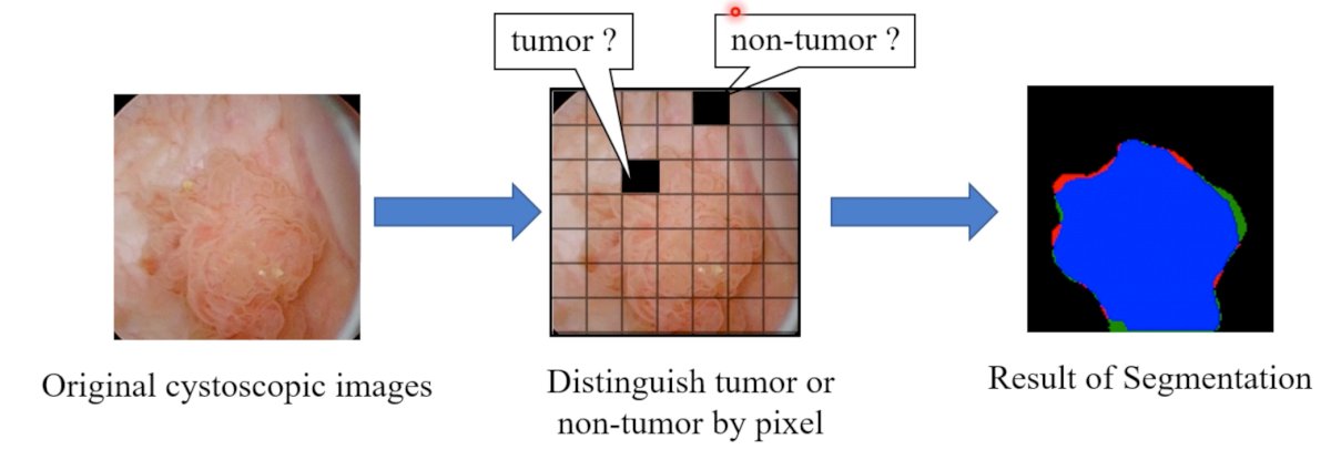
What is an AI segmentation system? Segmentation is an objective, reproducible system that can display the tumor regions in segmented pixels. As demonstrated below, the original cystoscopic image is segmented into pixels and each pixel is noted as having tumor or not. The results of segmentation are collated via an image processing system.
The next question is: How do you create RCEI? Color digital images are made up of three channels (red, green, and blue channels). The red channel image is subsequently enhanced and the RCE image is created.

The objective of this study was to evaluate whether RCEI improve the segmentation accuracy of cystoscopic images compared to conventional white light images. To that end, the authors retrospectively collected bladder tumor cystoscopic images among patients who underwent a TURBT between April 2014 and December 2019.
The images were divided into training and testing data. They constructed a segmentation system based on U-Net and implemented the image processing system to automatically create RCEIs. The investigators next compared two training methods:
- Training 1 using only cystoscopic images
- Training 2 using RCEI
The authors next compared the accuracies of the two training approaches.

The investigators evaluated pixel-wise sensitivity, positive predictive value, and IoU, which represents the extent of overlap between the area predicted by AI and the ground truth image.

Of the 1,790 images obtained, 900 were of Ta and only 116 were Tis tumors. The RCEI-based training outperformed cystoscopic image-based training with regards to:
- Pixel-wise sensitivity: 88% versus 85%
- Pixel-wise PPV: 87% versus 85%
- IoU: 77% versus 73%
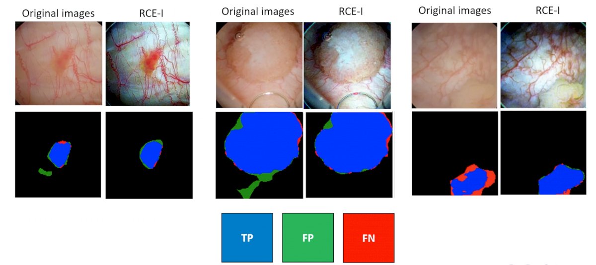
The authors concluded that RCEIs could improve segmentation accuracies. Although NBI uses blue and green wavelengths, RCEIs emphasize the red channel component and has the possibility to be used as a new diagnostic system that is different to NBI. RCEI-based AI can display the tumor regions as if they were PDD and has the ability to be an objective, reproducible system that can be repeatedly referenced during cystoscopy.`
Presented by: Dr. Jun Mutaguchi, MD, Department of Urology, Kyushu University Hospital, Fukuoka, Japan
Written by: Rashid K. Sayyid, MD, MSc – Society of Urologic Oncology (SUO) Clinical Fellow at The University of Toronto, @rksayyid on Twitter during the 2023 European Association of Urology (EAU) Annual Meeting, Milan, IT, Fri, Mar 10 – Mon, Mar 13, 2023.