

Our recently published study in the Journal of Vascular and Interventional Radiology focuses on one of the most practical aspects of renal cryoablation: the relationship between the visible iceball margin and subsequent local treatment failure.1
The study included 575 biopsy-proven cT1a renal cell carcinomas treated with CT-guided percutaneous cryoablation at Aarhus University Hospital between 2015 and 2022. Overall, local treatment failure occurred in only 17 tumors, corresponding to a rate of 3.0%. For tumors ≤3 cm, the local treatment failure rate was 1.8%. These results support the conclusion that in an experienced high-volume center, CT-guided renal cryoablation can achieve excellent, durable local tumor control.
A key debate in renal tumor ablation is whether local failure is driven by tumor biology or by insufficient technical coverage. This distinction matters: biology-driven failure leaves little room for procedural improvement, whereas margin-driven failure makes planning, probe placement, and intraprocedural margin assessment central to better outcomes. Our results strongly support the latter interpretation.
Tumors with local treatment failure had substantially smaller minimum tumor-to-iceball margins than matched tumors with durable local control. In the matched analysis, the mean minimum treatment margin was 0.4 mm in tumors with local treatment failure compared with 7.1 mm in size- and histology-matched local controls. The predefined 5-mm clinical margin threshold performed very well, with an AUC of 0.97 and a prevalence-adjusted negative predictive value of 99.6%.
Most importantly, all in-zone local treatment failures occurred when the minimum treatment margin was <5 mm. In practical terms, when a recurrent or residual tumor appeared within the treated ablation zone, the initial visible iceball margin had been insufficient. This provides strong support for a simple procedural principle: for cT1a renal cell carcinoma, the operator should strive for a visible iceball margin of at least 5 mm whenever safely achievable.
Two cases of local tumor progression occurred outside the estimated ablation zone despite apparently sufficient margins. Both were located adjacent to Gerota’s fascia and may represent distinct clinical phenomena, such as tract-related tumor seeding, perinephric progression, or fat necrosis mimicking recurrence. These cases underline the need for careful interpretation of perinephric findings on follow-up imaging, but do not change the central finding that in-zone failure was strongly linked to insufficient margin.
From an interventional perspective, the study provides pragmatic guidance. Intraprocedural CT should confirm not just that the tumor lies inside the iceball, but that it is covered with an adequate margin in all relevant planes. If coverage appears insufficient, the operator can consider adjusting the probe configuration, extending the freeze, adding another cryoprobe, modifying the probe power, or improving the displacement of adjacent structures. The visible iceball is one of the major advantages of cryoablation, but its value depends on active assessment of the margin during the procedure.
The temporal pattern of failures is also important. The highest local treatment failure rate occurred in 2015–2016, during the transition from laparoscopic-assisted cryoablation to fully CT-guided percutaneous cryoablation at Aarhus University Hospital. This distinction matters since the local failure rate was considerably higher in the earlier laparoscopic-era Aarhus cohort, with 11% residual unablated tumor and 3% late local recurrence.2 In contrast, the present CT-guided cohort had an overall local treatment failure rate of 3.0%, and only 1.9% from 2017–2022, after the transition period.
This highlights a broader issue for guidelines and shared decision-making: historical ablation data often combine data from different eras, techniques, tumor sizes, imaging modalities, and ablation approaches.3 Such legacy data remain important, but should not be interpreted as directly equivalent to contemporary CT-guided percutaneous cryoablation. Modern procedures benefit from improved CT systems, multiplanar navigation, systematic probe planning, hydrodissection and displacement techniques, and better visualization of the iceball–tumor relationship.
Our findings should therefore be interpreted in the context of technical maturation and margin adequacy. Although the temporal association cannot prove causality, it supports a clinically meaningful learning curve and suggests that reliance on legacy ablation data may overestimate the risk of local failure after modern percutaneous cryoablation of well-selected cT1a tumors.
The findings are also highly relevant for patient counseling. Patients treated with renal cryoablation often want to know how confident the treatment team can be that the tumor has been completely destroyed. This concern should not be underestimated. A recent Danish multicenter study by Bak and colleagues showed that fear of cancer recurrence is not confined to patients managed with active surveillance.4 Patients treated with cryoablation had comparable levels of fear of recurrence, and 28% of cryoablation patients reported scores consistent with clinical fear of cancer recurrence.
Even after minimally invasive treatment, some patients continue to worry that the cancer may return, particularly around follow-up imaging. In this context, our study provides a concrete way to meet that fear with evidence. If an adequate visible iceball margin of at least 5 mm has been documented in a cT1a tumor, and especially if early follow-up imaging confirms complete treatment, patients can be told that the subsequent risk of local recurrence appears to be extremely low.
This changes the counseling message from a general reassurance “the procedure went well” to a more specific and evidence-based statement: “We documented an adequate treatment margin, and in our data this was associated with a very low risk of local failure.” For many patients, that distinction may matter.
Looking ahead, improved intraprocedural imaging may further strengthen renal cryoablation. Spectral CT, virtual monoenergetic imaging, electron-density imaging, and deep-learning CT reconstruction may improve iceball conspicuity and confidence in margin assessment.5 In the longer term, CT thermometry may allow ablation monitoring to move beyond geometric iceball visualization toward more direct assessment of thermal injury.
In summary, our study suggests that local failure after CT-guided cryoablation of cT1a renal cell carcinoma is strongly associated with insufficient visible iceball margin. A 5-mm margin threshold provides practical guidance for interventionists, supports procedural quality assurance, and offers concrete reassurance for patients after treatment. In a high-volume center, particularly after procedural maturation, CT-guided renal cryoablation can achieve excellent local tumor control and should be considered an important nephron-sparing treatment option within modern multidisciplinary kidney cancer care.
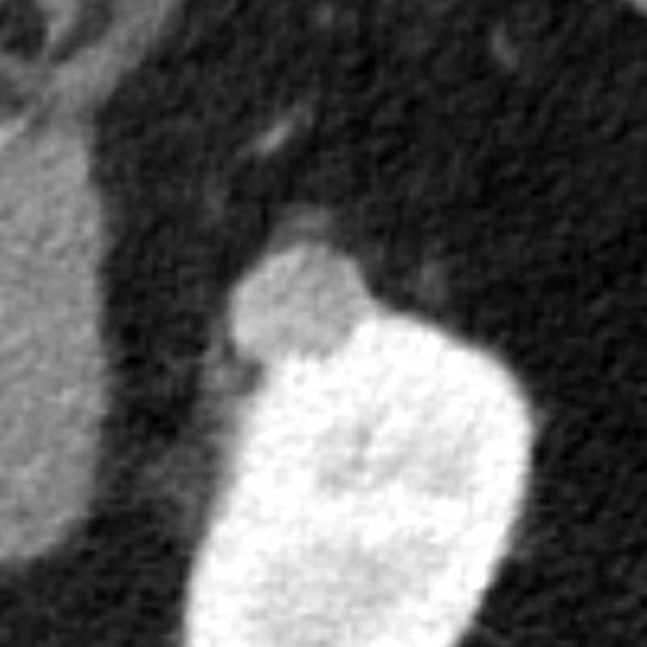
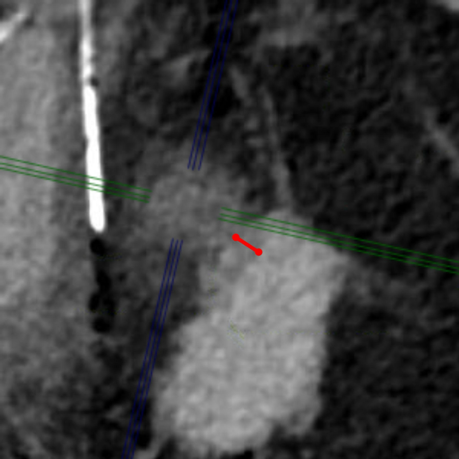
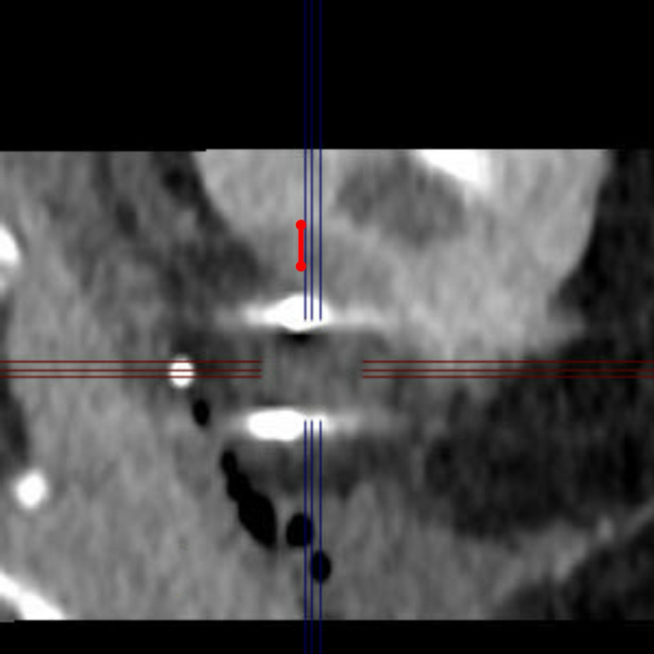
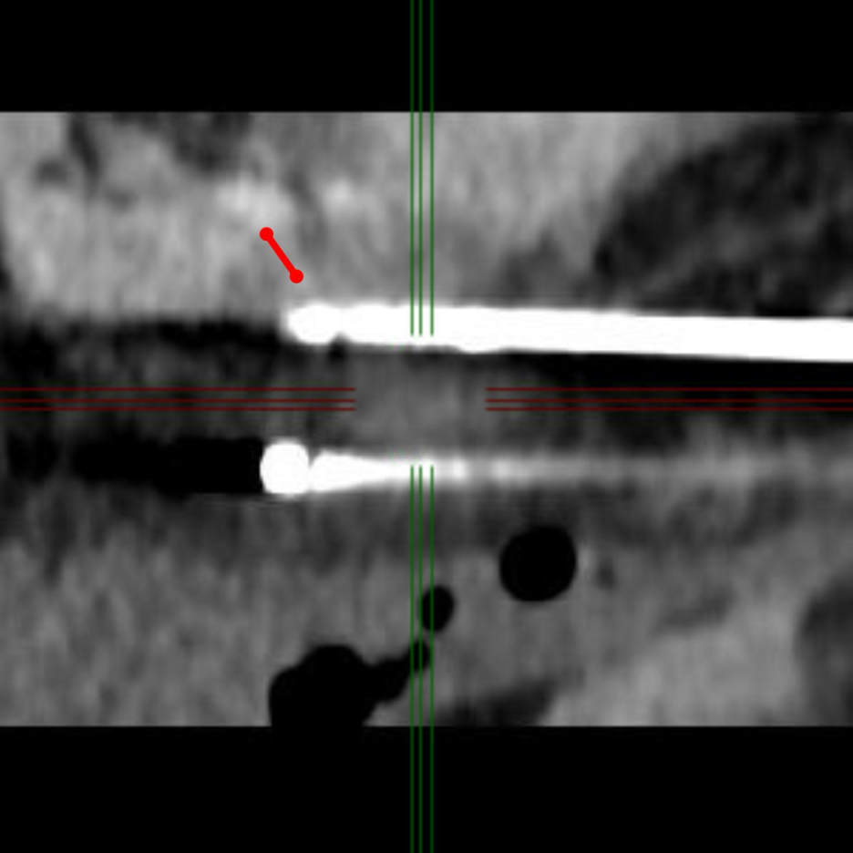
Figure. Intraprocedural CT assessment of iceball margin during renal cryoablation. (A) Contrast-enhanced planning CT demonstrates an enhancing exophytic renal tumor in the left kidney selected for CT-guided cryoablation. (B–D) Intraprocedural CT images obtained during cryoablation show cryoprobe placement and progressive iceball formation around the tumor. The red calipers illustrate measurement of the minimum visible tumor-to-iceball margin, defined as the shortest distance between the tumor boundary and the outer margin of the iceball.
Adequate intraprocedural margin assessment is central to ensuring complete tumor coverage, with a visible iceball margin of at least 5 mm recommended whenever safely achievable.
Written by:
- Jens Borgbjerg, MD, PhD, Department of Radiology, Akershus University Hospital, Norway
- Mia Gebauer Madsen, MD, PhD, Department of Urology, Aarhus University Hospital, Denmark
- Ole Graumann, MD, PhD, Department of Radiology, Aarhus University Hospital, Denmark
References:
- Borgbjerg J, Madsen MG, Al-Mashhadi R, Andersen G, Brandt SB, Junker T, et al. Iceball Margin <5 mm Is Associated with Local Failure after CT-guided Cryoablation of cT1a Renal Cell Carcinoma. J Vasc Interv Radiol. 2026; 108905.
- Nielsen TK, Vedel PF, Borgbjerg J, Andersen G, Borre M. Renal cryoablation: five- and 10-year survival outcomes in patients with biopsy-proven renal cell carcinoma. Scand J Urol. 2020;54: 408–412.
- Stamper M, Collins J, Arellano RS, DeSanto M, Deem S, Annie F, et al. Long-term Outcomes of Partial Nephrectomy Versus Percutaneous Ablation for Renal Cell Carcinoma: A Propensity-Matched Analysis. Urology. 2025;206: 66–73.
- Bak R, Junker T, Jensen JB, Pelant T, Haase RN, Zachariae R, et al. A comparative analysis of fear of cancer recurrence in patients with small renal masses: Active surveillance versus cryoablation. Acta Oncol. 2024;63: 573–579.
- Borgbjerg J, Rafdal H, Rasmussen GE, Johansen TJ, Svendsen ED, Graumann O. Improved Ice Ball Visualization during CT-Guided Cryoablation of Renal Tumors Using Dual-Energy CT Virtual Monoenergetic and Electron Density Images. J Vasc Interv Radiol. 2025;36: 716–719.