

The typical complex robotic partial nephrectomy according to the panel is a 66-year-old African American female with a right 5.5 cm renal mass, which is posterior, mid/upper pole, partially cystic, as well as endophytic and hilar (RENAL score 10ph). Her baseline eGFR is 39, with type 2 diabetes, hypertension, obesity (BMI 43) and with a prior open cholecystectomy. A biopsy of this mass demonstrates clear cell RG, Fuhrman grade 1. This complex case is managed with a transperitoneal approach with renal hypothermia with ice slush via an extra trocar. She has an uneventful post-operative course and is discharged on post-operative day #2 with a Cr spike to 2.46 (1.67 pre-operatively), which levels off at 2.1 (eGFR 30). The final pathology is clear cell RCC, Fuhrman grade II, pT1b with negative margins. With increasing comorbidities and obesity rates amongst our patients, we are going to be increasingly seeing these types of index complex cases.
Dr. Michael Stifelman then presented a complex case utilizing a retroperitoneal approach. This patient was 69 years-old with a Bosniak II cyst in 2012, eGFR >60, however follow-up ultrasound demonstrated this cyst was now complex. A subsequent CT scan demonstrated a 4.2 cm Bosniak IV cyst and the patient had a history of prior left colectomy for diverticulitis. For these cases, Dr. Stifelman uses the Xi platform to take advantage of the narrow arms, closer ports and side docking. Additionally, he places the patient in a complete lateral (90 degree) decubitus position to easily access the retroperitoneum. As he notes, even in super morbid obese patients (ie. BMI >60) the adipose tissue typically is anterior with limited fat deposition in the retroperitoneum. Here is his port placement for a robotic retroperitoneal partial nephrectomy:
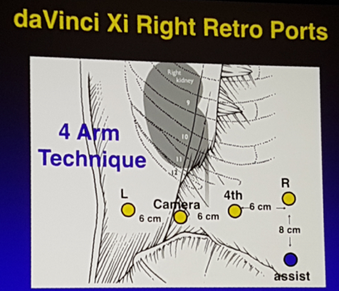
For access, Dr. Stifelman prefers the da Vinci Hasson port. This patient ultimately underwent an uncomplicated procedure with a warm ischemia time of 19 minutes, and was discharged on post-operative day #1. Final pathology demonstrated a 4.0 cm cystic RCC, Fuhrman grade 3 with negative margins.
Dr. Monish Aron then discussed the complex robotic partial nephrectomy for cT1b/T2 renal masses. A case was presented of a 57-year-old female with an 8 cm right renal mass on CT scan, in addition to a staghorn calculus in the left kidney. She had a medical history consisting of hypertension, nephrolithiasis, metabolic syndrome, and a Cr of 0.97 (eGFR 65). A functional renogram demonstrated 62% function on the right and 38% function on the left, and a needle biopsy confirmed a clear-cell RCC. This patient underwent a successful transperitoneal robotic partial nephrectomy and had an uncomplicated post-operative course. As Dr. Aron mentions, with the increasingly comorbid population we will be increasingly seeing these patients with complex masses that will benefit from a meticulous and safe complex robotic partial nephrectomy approach.
Dr. Jihad Kaouk then concluded this session by presenting surgical hints for managing renal sinus fat invasion during a robotic-assisted partial nephrectomy. He started by highlighting that the renal sinus is the fatty component located within the confines of the kidney not delineated from the renal cortex by a fibrous capsule. A case he presented was of a patient with a 4.0 cm left renal mass, endophytic and abutting/invading the renal sinus fat (RENAL score 9p). Dr. Kaouk highlighted during a video presentation of this patient’s case that it is important to be methodical with clear visualization in order to deftly excise these complex lesions invading the sinus fat. He also noted that he will often use absorbable clips to control bleeding, particularly when he is excising the tumor deep in the sinus fat (also reminding us that non-absorbable clips should not be used secondary to the proximity to the collecting system).
Dr. Kaouk emphasized a very recently published study utilizing the SEER database to assess the impact of upstaging patients to pT3a. For this study, from 1998-2013, 28,854 pT1-pT3aN0M0 kidney cancer patients were identified as having undergone a partial nephrectomy [1]. Among these patients, the estimated proportion up-staging to pT3a was 4.2%, 9.5%, and 19.5% for cT1a, cT1b, and cT2, respectively. Overall survival (OS) was worse for tumors up-staged from cT1a to pT3a, but not for cT1b or cT2 tumors. Up-staged pT3a tumors across all stage strata demonstrated worse CSS, with worse survival for larger tumors. Finally, for tumors <4 cm, univariable models showed improvement in OS and CSS for patients receiving a partial nephrectomy for pT3a tumors (up-staged from cT1a) relative to those receiving a radical nephrectomy for similarly sized pT3a tumors.
Dr. Kaouk provided several important take-home messages:
- Robotic partial nephrectomy is safe and feasible for tumors invading the sinus fat in the hands of experienced robotic surgeons
- pT3a up-staging occurs in roughly 4% of cT1a tumors and up to 20% for cT2c tumors
- Up-staged patients have worse survival than counterparts with concordant pathology
- Up-staged tumors undergoing partial nephrectomy represent a low-risk pT3a subset
Presented By: Monish Aron, University of Southern California, Los Angeles, CA; Jihad Kaouk, Cleveland Clinic Foundation, Cleveland, OH; Michael Stifelman, Hackensack University, Hackensack, NJ
Written By: Zachary Klaassen, MD, Urologic Oncology Fellow, University of Toronto, Princess Margaret Cancer Centre, @zklaassen_md ,at the 2018 North American Robotic Urology Symposium, February 16-17, 2018 - Las Vegas, NV
References:
- Srivastava A, Patel HD, Joice GA, et al. Incidence of T3a up-staging and survival after partial nephrectomy: Size-stratified rates and implications for prognosis. Urol Onc 2018;36(1):12.e7-e12.