(UroToday.com) The 39th World Congress of Endourology and Uro-Technology included a kidney imaging and thermal ablation session featuring work from Dr. Christopher Staniorski and colleagues presenting results of their study assessing the accuracy of radiology reports of ureteral stone size from computed tomography (CT) scans obtained in the emergency department (ED). Currently, nephrolithiasis and renal colic account for over 2 million ED visits per year. While a CT scan can demonstrate the presence of obstructing ureteral stones, the measurement of these stones is critical. The size of the stone determines the plan of care, including the prediction of stone passage and the necessity of intervention. The omission of stone measurement dimensions can therefore impact proper measurement.
In this study, researchers sought to understand the differences in practice patterns in reporting stone dimensions on CT scans between radiologists and urologists, understand differences in maximal diameter based on the number of stone dimensions measured, and any resulting changes in patient course due to these differences. A retrospective chart review was conducted at a single tertiary center, in which adults who presented to the ED with a single unilateral ureteral stone were included in the study. Charts were reviewed for radiologist reports of stone dimensions, followed by a retrospective measurement by a urologist who calculated the stone in the anterior-posterior (AP), lateral, and craniocaudal (CC) dimensions.
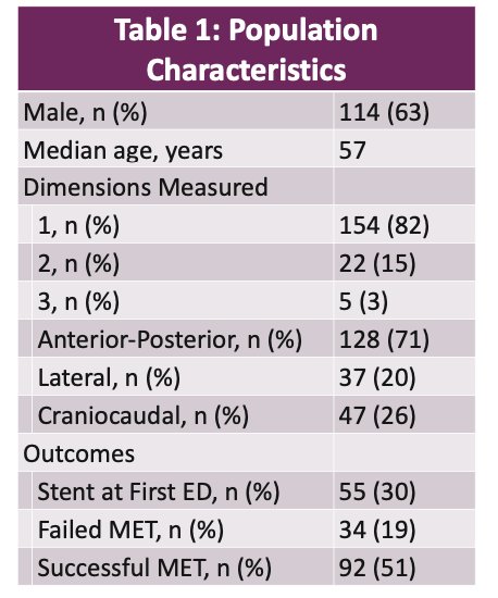
Table 1 displays a summary of the 181 patients included in the study. Notably, only 3% of all radiology reports included all 3 dimensions. In considering all dimensions, median stone measurement by urologists was significantly greater than measurement by radiologists. However, in the cases when the radiologist reported the craniocaudal dimension, there was no significant difference in stone measurement. In this subpopulation, stone size was overall significantly underreported in radiology reports.
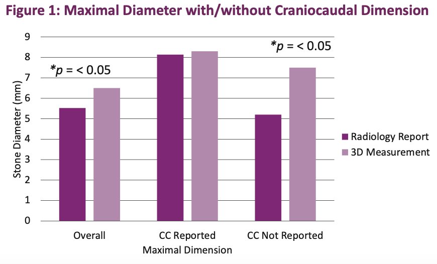
Figure 1 demonstrates this effect and points to the conclusion that while CC dimension is frequently omitted, its inclusion often reconciles discrepancies in stone size reports.
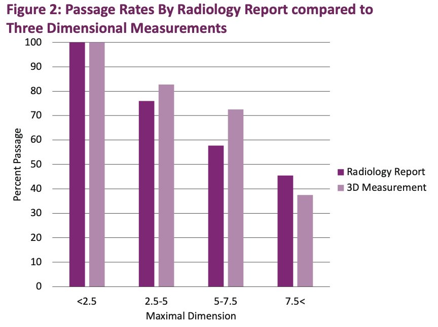
In figure 2, the question of this phenomenon’s impact on clinical outcomes is explored. Both the radiologist report and 3D measurements by urologists both correlated with stone passage. Therefore, the prognostic importance of these discrepancies is unclear.
In conclusion, Dr. Staniorski emphasized that omission of the craniocaudal dimension is not only frequently omitted, but often measures the largest dimension when all three are considered. This data, therefore, underscores the importance of personal imaging review by the managing urologist.
During discussion, a comment was made on the importance of this work and the impact of the radiology report on not only the prediction of passage rates, but also the prescription of alpha blockers. While this is often done indiscriminately, a urologist reviewing the imaging allows for greater knowledge for making this decision to prescribe Flomax. One of the moderators, Dr. Landman, proceeded to ask a question about stratifying the data between medical centers to see a difference in quality of reads. He went on to state that the comparison of outside reports to reports done at the institution may be interesting to understand, thus offering an intriguing avenue for future study. A final commenter discussed the importance of sharing this data with emergency department physicians and radiologists to foster interdisciplinary collaboration and better patient management.
Presented by: Christopher Staniorski, MD – University of Pittsburgh Medical Center
Written by: Kelvin Vo, BS, Assistant Research Specialist, Department of Urology, University of California Irvine, @kelvinvouci on Twitter during the 39th World Congress of Endo urology and Uro-Technology (WCET), Oct 1 - 4, 2022, San Diego, California.
References:
- C Staniorski, D Pelzman, M Semins. Accuracy of Radiology Reports of Ureteral Stone Size in the Emergency Department [abstract]. In: 39th World Congress of Endourology and Technology, October 1-4, 2022, San Diego, CA