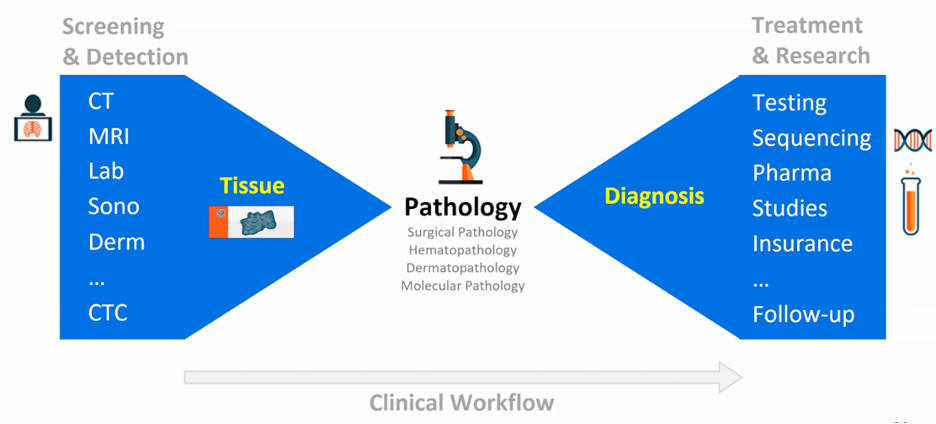
According to Dr. Fuchs, pathologists are under stress and are headed for a work crisis with an increase in new cancer cases per pathologist from 2007 to 2017, and a dwindling workforce during the same time period. Additional work has shown that the percentage of pathologists experiencing adverse events increases significantly with a workload greater than 39 hours per week. These statistics have opened the door for computational pathology. A state of the art dataset in pathology would be tiny (~400 slides), and very well curated. However, the clinical reality is that clinical practice is messy, diverse, and surprising. Dr. Fuchs notes that there are several aspects that it takes to go beyond this current state:
- Machine learning: new ways to learn from data at a petabyte scale
- Data: real-world, clinically relevant datasets at scale
- Domain experts: pathologists and computer scientists working in tandem
- Computation: high-powered computers for efficient deep learning at scale
The classical, supervised model of grading slides is pixel-level annotation, whereas weakly supervised learning is image-level annotation, which is a binary label for the whole slide from the pathology report. However, a prostate cancer lesion may be small and thus there are inherent challenges.
In work from Dr. Fuchs’ group to overcome the problem of being hindered by the need for large manually annotated datasets for the development of decision support systems for pathology and their deployment in clinical practice, they developed a multiple-instance learning-based deep learning system that uses only the reported diagnoses as labels for training, thereby avoiding expensive and time-consuming pixel-wise manual annotations.1 This framework was evaluated at scale on a dataset of 44,732 whole slide images from 15,187 patients without any form of data curation. This included tests on prostate cancer, basal cell carcinoma, and breast cancer metastases to axillary lymph nodes, which resulted in areas under the curve above 0.98 for all cancer types. According to Dr. Fuchs, this clinical application would allow pathologists to exclude 65-75% of slides while retaining 100% sensitivity. As follows is the receiver operating characteristic (ROC) curve for prostate cancer:
A recently published study, also from Dr. Fuchs’ group, assessed the impact of artificial intelligence systems on pathologic diagnoses.2 They investigated how pathologists interact with Paige Prostate Alpha, a state-of-the-art prostate cancer detection system, in whole slide images of prostate needle core biopsies stained with hematoxylin and eosin. Three board-certified pathologists assessed 304 anonymized prostate needle core biopsy whole slide images in eight hours. After four weeks, pathologists were tasked with re-reviewing each whole slide image with the aid of Paige Prostate Alpha. Against the ground truth, the pathologists and Paige Prostate Alpha were measured: without Paige Prostate Alpha, pathologists had an average sensitivity of 74% and an average specificity of 97%, whereas, with Paige Prostate Alpha, the average sensitivity for pathologists significantly increased to 90% with no statistically significant change in specificity:
Dr. Fuchs concluded with several points regarding the potential impact of artificial intelligence on patients:
- Better care at a lower cost
- 100% quality assurance
- Molecular classification
- Prediction of cancer progression
- Predicting treatment response
Presented by: Thomas J. Fuchs, MD, PhD, Department of Pathology, Memorial Sloan Kettering Cancer Center, Associate Professor, Weill Cornell Graduate School for Medical Sciences, Director, The Warren Alpert Center for Digital and Computational Pathology, New York, New York
Written by: Zachary Klaassen, MD, MSc, Assistant Professor of Urology, Georgia Cancer Center, Augusta University/Medical College of Georgia, Augusta, Georgia, Twitter: @zklaassen_md at the 2020 Society of Urologic Oncology Annual Meeting – December 2-5, 2020 – Washington, DC
References:
1. Campanella, Gabriele, Matthew G. Hanna, Luke Geneslaw, Allen Miraflor, Vitor Werneck Krauss Silva, Klaus J. Busam, Edi Brogi, Victor E. Reuter, David S. Klimstra, and Thomas J. Fuchs. "Clinical-grade computational pathology using weakly supervised deep learning on whole slide images." Nature medicine 25, no. 8 (2019): 1301-1309.
2. Raciti, Patricia, Jillian Sue, Rodrigo Ceballos, Ran Godrich, Jeremy D. Kunz, Supriya Kapur, Victor Reuter et al. "Novel artificial intelligence system increases the detection of prostate cancer in whole slide images of core needle biopsies." Modern Pathology (2020): 1-9.