(UroToday.com) The 2025 SNMMI annual meeting featured a prostate cancer and molecular imaging session and a presentation by Dr. Yujia Li discussing a head-to-head comparison of mpMRI, PSMA, and gastrin-releasing peptide receptor (GRPR) PET/CT for local staging of primary prostate cancer. Optimal therapeutic options for prostate cancer rely on the accurate assessment of the local extent, localization, and aggressiveness of the primary tumor. Advanced imaging plays a crucial role in the initial evaluation of prostate cancer, with enhanced sensitivity potentially leading to stage migration. PSMA and GRPR are overexpressed in different patterns in prostate cancer. While PSMA PET/CT has been assessed for local staging, the feasibility of GRPR PET/CT in this context has not been explored, nor has its comparison to mpMRI or PSMA PET/CT. Additionally, the combined use of PET/CT + mpMRI has been inadequately investigated. This study, presented at the SNMMI 2025 annual meeting, reports an exploratory analysis from a prospective, single-center, single-arm trial, comparing the efficacy of these imaging modalities in identifying prostate cancer local stage.
Patients with intermediate-/high-risk prostate cancer who underwent mpMRI, PSMA, and GRPR PET/CT before radical prostatectomy in a prospective paired clinical trial were included. Each modality was interpreted by two independent readers unaware of clinical or other imaging findings, and histopathology from radical prostatectomy served as the gold standard. Imaging T staging was assessed, in addition to bilateral intraprostatic disease (T2c), extraprostatic extension (T3a), and seminal vesicle invasion (T3b). VOI-assisted analysis was used for equivocal findings on PET/CT images. The index lesion was defined as the most clinically significant lesion (highest tumor stage, PET SUVmax, or MRI PI-RADS). PET/CT + mpMRI findings were obtained by combining the final results from two modalities. Accuracy in detecting the index lesion, extraprostatic extension, seminal vesicle invasion, and T staging was summarized.
The analysis included 81 treatment-naïve prostate cancer men (40 intermediate-risk [49%], and 41 high-risk [51%]). Prostate adenocarcinomas were found in 81 (100%) cases from radical prostatectomy, and 10 (12%) had a mixture with unconventional pathological patterns. PSMA PET/CT showed greater accuracy in local staging than GRPR PET/CT (56% versus 36%, p = 0.011), but was not superior to mpMRI (56% versus 41% p = 0.073). However, PSMA PET/CT was superior to both modalities in the pure adenocarcinoma subgroup (58% versus 39% and 34%, p = 0.029 and 0.005). PSMA PET/CT was also superior to mpMRI for bilateral intraprostatic disease identification (72% versus 54%, p = 0.024), and superior to GRPR PET/CT for seminal vesicle invasion identification (93% versus 85%, p = 0.039). Combining PSMA PET/CT with mpMRI demonstrated superior accuracy to mpMRI alone (60% versus 41%, p = 0.002), and GRPR PET/CT with mpMRI was superior to GRPR PET/CT alone (49% versus 36%, p = 0.035). The combination of PET/CT + mpMRI detected more index lesions (93% and 89% versus 77% p < 0.001 and p = 0.002) and identified bilateral intraprostatic disease with higher accuracies (74% and 67% versus 54%, p = 0.002 and 0.041) compared to mpMRI alone. The following images show mpMRI, PSMA, and GRPR PET/CT comparing various whole mount pathology ISUP 2 lesions:
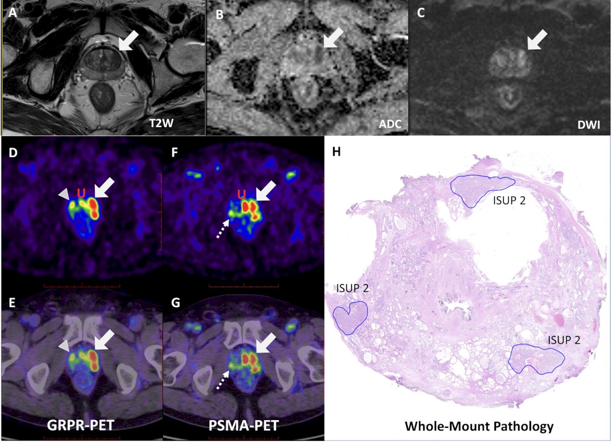
The following images show a GRPR PET/CT SUVmax 23.1 lesion corresponding to a whole mount pathology ISUP 2 lesion:

Dr. Li concluded this presentation discussing a head-to-head comparison of mpMRI, PSMA, and GRPR PET/CT for local staging of primary prostate cancer with the following take home points:
- In patients with intermediate- and high-risk prostate cancer, PSMA PET/CT exhibited the highest local staging accuracy among the three modalities, with statistical superiority in cases with pure adenocarcinoma pathology
- This advantage was primarily due to better identification of bilateral intraprostatic disease, followed by index lesions and seminal vesicle invasion
- Underestimation of pathological local staging by the three modalities is not uncommon
- Combining mpMRI with PET imaging enhances the local staging accuracy compared to mpMRI alone, allowing for the more accurate identification of index lesions and bilateral intraprostatic disease
- This improvement may be clinically significant for more precise targeted biopsy guidance and local treatment planning
Presented by: Yujia Li, Institute of Computing Technology, Chinese Academy of Sciences, Beijing, China
Written by: Zachary Klaassen, MD, MSc – Urologic Oncologist, Associate Professor of Urology, Georgia Cancer Center, Wellstar MCG Health, @zklaassen_md on Twitter during the 2025 Society of Nuclear Medicine and Molecular Imaging (SNMMI) Annual Meeting, New Orleans, LA, June 21st – 24th, 2025