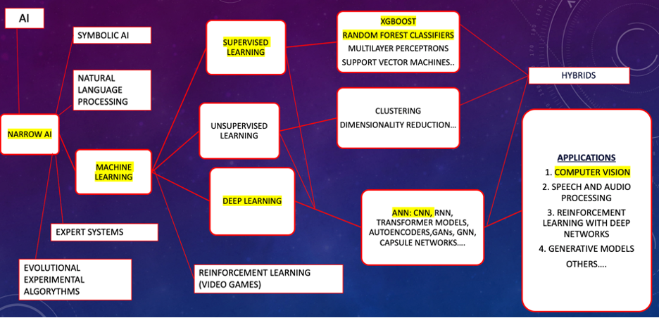
She then discussed random forest classifiers, which are an ensemble method that combines multiple decision trees, while artificial neuronal networks are non-ensemble non-linear models inspired by neurons in the decision-making process. Next, Dr. Bird discussed an equation used for artificial intelligence algorithm development. By leveraging this organized data, artificial intelligence algorithms can be developed more effectively, enabling precise patterns recognition and predictive modeling:

Factors are grouped into phenotype, genotype, epigenetics, and environment, which are the key aspects to the phenotypic variance equation:

Regarding artificial intelligence used for prostate cancer diagnosis, there are three commercial artificial intelligence software platforms, two from the United States and one from Japan. The first two (Paige Prostate Alpha and the Japanese platform), use digital reading of scanned pathological slides and predict prostate cancer with a sensitivity of 90-100%, however, specificity is not improved with this methodology. IBEX combines heat maps to identify prostate cancer with improved specificity of 97%:

A new technique using stimulated Raman scattering microscopy uses CNN-based artificial intelligence algorithms to accurately recognize the molecular vibrational fingerprint of cancer versus non-cancerous tissue, in addition to a superior specificity of 96%. This is done without staining and is processed within 2 minutes, making this a potential cutting-edge technology, which could change the current pathology interpretation paradigm:

Dr. Bird then discussed the role of artificial intelligence in ultrasound and MRI of the prostate. Prostate volume assistance uses convolutional neuronal networks to measure prostate volume, with no significant difference between human and PVA measurements. However, both are equally different from MRI volumes, consistent with an MRI’s tendency to overestimate prostate volume. A second artificial algorithm using pathological and MRI imaging were used to train this model, to predict ultrasound prostate cancer lesions, and outperforming the human reader. A third model uses multiparametric ultrasound images to train an artificial intelligence ultrasound-based algorithm, using shear wave elastography to visualize prostate lesions, in addition to using gray scale to illustrate hypoechoic lesions. Again, shear wave elastography demonstrates benign tissue in the transitional zone, and using computational algorithms, this model can predict prostate cancer lesions with an AUROC of 0.60:

There are multiple artificial intelligence software available for MRI segmentation. Two commercially used software include the Quatib European based artificial intelligence, acquired by RadNed, which uses CNN algorithms to segment and localize lesions, as well as design PI-RADS reading scores. Prosta ID uses 3D voxel texture CNN architecture to segment prostate lesions:

A third artificial intelligence software to segment the prostate is the artificial intelligence platform Protégé by Mims GE Health:

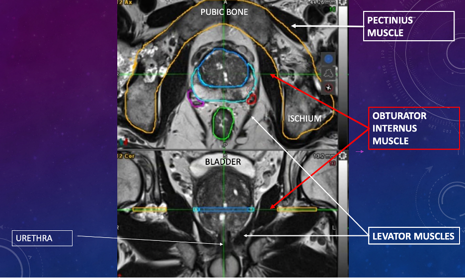
Using multiparametric MRI, artificial intelligence can be trained to auto-contour other areas, including neurovascular bundles, in addition to muscular structures, such as the obturator internus and levator muscles, and most importantly the apical prostate and urethral complex:

In work from Dr. Bird’s group, as the artificial intelligence platform Protégé was developed, it yielded a Dice coefficient of more than 0.75 compared to a board certified urologists and radiologists. The average time to produce the images was more time efficient when compared to human produced contours.1
 A limitation of artificial intelligence applications in MRI segmentation is the current PI-RADS system used to train machine learning models secondary to the high inter- and intra-reader variability of the PI-RADS scoring system. Other MRI reading methodologies are being developed, such as quantitative ADC mapping analysis, but these models are still being investigated and will need further data to validate the current findings. In addition, metallic objects, such as hip prostheses and Urolift clips, can cause significant MRI distortion, making DWI, ADC, and DCE series mapping not amenable to interpretation.
With regards to ablative technologies, TULSA PRO has 10 parallel high frequency ultrasound beams in a closed-loop, heating to ablative temperatures of 55°C. This energy is delivered with real time MR thermometry to assure adequate temperature conditions for accurate energy delivery for tissue ablation:
Artificial intelligence technology is used in the planning part of the procedure, making this aspect more efficient. The five year outcomes for TULSA demonstrated a failure rate of 21% at 5 years, with 92% and 87% continence and potency rates, respective.
Avenda Health artificial intelligence segmentation of tumors was designed to address the underestimation of margins to improve focal therapy accuracy for Focal One Robotic HIFU. This technology noted improved margins of 97% versus 38% for human derived segmentation:
Ultrasound-guided Focal One robotic-assisted HIFU has implemented artificial intelligence-assisted localization of the tumor for improved treatment delivery. Other ultrasound-guided ablative technology, such as cryotherapy, IRE, and transperineal laser ablation of the prostate do not have artificial intelligence integration into their software, however these technologies can adapt to any MRI ultrasound fusion software.
To date, the use of artificial intelligence in the future of autonomous robotic system development is limited due to the regulatory and ethical implications. Several robotic systems are available, such as Dexter from Switzerland and Hinotori from Japan:
In the United States, the Da Vinci robotic system uses motion scaling and tremor reduction, in addition to 3D visualization during prostatectomies, to aid the surgeon. With artificial intelligence driving augmented reality identification of prostatic lesions, we are able to identify lesions in advanced pT3 disease, allowing the use of nerve-sparing surgery without compromising surgical margins:
Using automated surgical video analysis, previous studies have achieved a concordance rate of 93% between artificial intelligence and humans pertaining to documentation that can be used, not only in surgical training and education, but also in medical billing:2

Discussing radiation and artificial intelligence, RaySearch’s RayStation uses artificial intelligence algorithms to adjust the amount of radiation as the tissue changes in shape and size. A commonly used artificial intelligence software in radiation oncology is ArteraAI, which uses clinical and pathological information to assess a risk score in patients and the benefit of using ADT in patients concurrently undergoing radiation:3

Finally, with regards to progression, OncoPredict is a non-commercial artificial intelligence software used in research that uses clinical data (including genetic and epigenetic data) to calculate cancer progression, as well as the phenotype that may benefit from certain treatments:

Using clinical and pathological data, Kwon et al.4 demonstrated that PSA is the most important factor for predicting extracapsular extension, with an AUROC of 0.81:
Additionally, T2W images have been used to develop an algorithm to predict extracapsular extension with an AUROC of 0.92 for assessing image quality, and an AUROC of 0.88 for the presence of extracapsular extensions.5
Dr. Bird concluded her presentation discussing artificial intelligence applications in prostate cancer diagnosis and treatment by emphasizing that currently there is extensive artificial intelligence work being done for phenotype, moderate artificial intelligence work for genotype, limited artificial intelligence work for epigenetics, and negligible artificial intelligence work for environment:
Presented by: Victoria Bird, MD, National Medical Association and Research, Gainesville, FL
Written by: Zachary Klaassen, MD, MSc – Urologic Oncologist, Associate Professor of Urology, Georgia Cancer Center, Wellstar MCG Health, @zklaassen_md on Twitter during the Southeastern Section of the American Urological Association (SESAUA) 2025 Annual Meeting, Nashville, TN, Wed, Mar 12 – Sat, Mar 15, 2025.
References:
- Shori P, Kim JH, Carey RI, et al. PD36-12 Artificial intelligence as a time-efficient and quality tool for contouring in prostate cancer diagnostics. J Urol. 2024 May;211(5S):e797
- Khanna A, Antolin A, Bar O, et al. Automated Identification of Key Steps in Robotic-Assisted Radical Prostatectomy Using Artificial Intelligence. J Urol. 2024 Apr 1 [cited 2024 Nov;211(4):575–584.
- Spratt DE, Tang S, Sun Y, et al. Artificial Intelligence Predictive Model for Hormone Therapy Use in Prostate Cancer. NEJM Evid 2023;2(8).
- Kwong JCC, Khondker A, Tran C, et al. Explainable artificial intelligence to predict the risk of side-specific extraprostatic extension in pre-prostatectomy patients. Can Urol Assoc J. 2022 Jun;16(6):213-221.
- Saikali S, Khosravi P, Gamal A, et al. MP07-02 Detection of Prostate Cancer Extracapsular Extension with MRI Using Deep Learning Methods. J Urol. 2024 May;211(5S):e105.