(UroToday.com) The 2025 South Central AUA annual meeting included a session on kidney cancer, featuring a presentation from Dr. Aaron Tverye discussing the utility of ultrasound compared to cross-sectional imaging in the follow-up of patients with renal cell carcinoma (RCC). RCC recurrence following surgical resection poses a significant challenge. Current surveillance guidelines following treatment of localized RCC from the AUA and the NCCN include imaging modalities such as CT, MRI, and ultrasound, with ultrasound recommended for the surveillance of patients deemed not to be high risk. Longer surveillance practice patterns are being used, given that approximately 30% of RCC recurrences are diagnosed after 5 years. To date, there is no definitive prospective evidence comparing the efficacy of imaging modalities, although retrospective data have reported ultrasound’s lower sensitivity for detecting smaller abdominal recurrences. This study aimed to evaluate the utility of ultrasound in detecting abdominal recurrence after definitive therapy for RCC and compare the detection rate to that of cross-sectional imaging.
This is a prospective clinical trial that has enrolled patients between January 2020 and December 2024. All patients underwent a partial or radical nephrectomy for RCC. Enrollment occurred when participants were identified to have intra-abdominal recurrence on cross-sectional imaging, either CT or MRI, based on routine surveillance. Once enrolled, patients with recurrences underwent an abdominal ultrasound at an outside facility performed by a blinded sonographer and read by an independent radiologist.
Fourteen patients were enrolled with a median age at recurrence of 72.5 years:

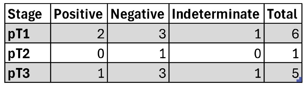
Seven patients (50%) underwent partial nephrectomy, and 7 patients (50%) underwent radical nephrectomy. Overall, 10 patients (71%) had clear cell RCC on final pathology. The median time to recurrence was 54 months following surgery (range 8-172). On average, an ultrasound was performed 3 months following the detection of recurrence. There were 4 (29%) ultrasound studies positively identifying intra-abdominal disease, while 8 (57%) were negative and 2 (14%) were indeterminate. Ultimately, 12 patients (86%) went on to undergo some form of secondary treatment, including four surgical resections and eight microwave ablations. False negatives were identified in patients with pT1-T3 disease:
Dr. Tverye concluded his presentation discussing the utility of ultrasound compared to cross-sectional imaging in the follow-up of patients with RCC with the following take home points:
- Ultrasound demonstrated poor sensitivity for detecting RCC recurrence and metastases, with false negatives seen in patients deemed low, intermediate, and high risk
- Though ultrasound is attractive due to its accessibility and lack of radiation exposure, future guidelines should reconsider the role of ultrasound in surveillance for patients of all risk categories
Presented by: Aaron Tverye, MD, University of Kansas Medical Center, Kansas City, MO
Written by: Zachary Klaassen, MD, MSc – Urologic Oncologist, Associate Professor of Urology, Georgia Cancer Center, Wellstar MCG Health, @zklaassen_md on Twitter during the 2025 South Central American Urological Association (AUA) Annual Meeting, Orlando, FL, Wed, Sept 10 – Sat, Sept 13, 2025.