(UroToday.com) The 2025 European Society of Medical Oncology (ESMO) Annual Congress held in Berlin, Germany was host to the session Mini Oral session 2: GU tumours, renal & urothelial. Dr. Alina T. Küper presented abstract 2597MO - [68Ga]Ga-DPI-4452 PET/CT for Staging of Patients with Clear Cell Renal Cell Carcinoma.
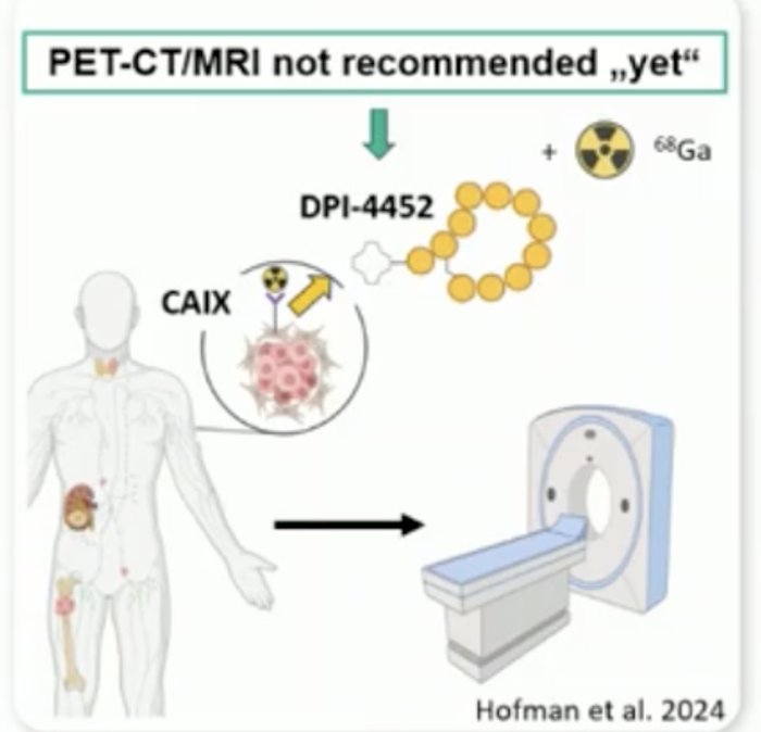
Dr. Küper and colleagues presented on the potential of [⁶⁸Ga]-DPI-4452 PET/CT for imaging CAIX expression in clear cell renal cell carcinoma (ccRCC), comparing its diagnostic accuracy and clinical impact with conventional CT. While CAIX is not yet a widely used imaging target, it is highly relevant given its overexpression in approximately 90% of ccRCC cases linked to VHL gene mutations and its role in acid–base regulation and tumor aggressiveness.
Since metastatic ccRCC carries a significantly worse prognosis, with OS dropping sharply after metastasis, accurate staging remains crucial. This ongoing study aims to assess detection efficiency across six anatomic regions and evaluate the theranostic potential of CAIX PET/CT through pre- and post-scan clinical questionnaires completed by urologic oncologists.
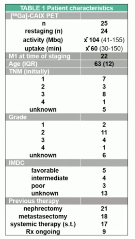
Dr. Küper presented the preliminary experience from the Essen group on the use of [⁶⁸Ga]-CAIX PET/CT in patients with clear cell renal cell carcinoma (ccRCC), highlighting its promising potential as both a diagnostic and theranostic tool. The study included 25 patients (median age 63 years), most of whom had advanced disease (22 with M1 at the time of staging) and prior systemic therapy (17 patients). The majority had undergone nephrectomy (21 patients), and the distribution of IMDC risk groups reflected a real-world advanced ccRCC population.
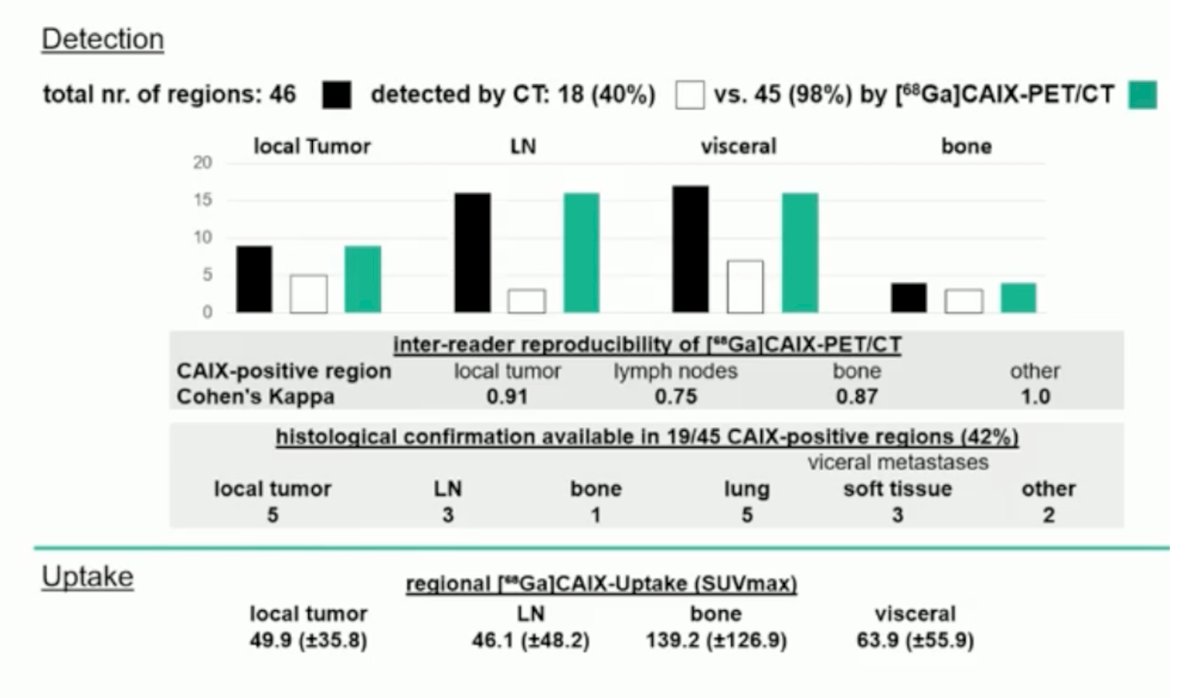
In terms of diagnostic performance, [⁶⁸Ga]-CAIX PET/CT detected 98% of evaluated lesions (45/46) compared to only 40% (18/46) detected by standard CT, demonstrating superior sensitivity across local tumor, lymph node, visceral, and bone regions. Inter-reader reproducibility was excellent, with Cohen’s kappa values of 0.91 for primary tumor, 0.75 for lymph nodes, and 0.87 for bone metastases. Histological confirmation was available in 42% (19/45) of CAIX-positive lesions, further supporting imaging accuracy. The highest tracer uptake (SUVmax) was observed in bone lesions (139.2 ±126.9), followed by visceral (63.9 ±55.9) and local tumor sites (49.9 ±35.8), indicating robust radiotracer affinity that may inform future radionuclide therapy development.
Clinically, the [⁶⁸Ga]-CAIX PET/CT findings had a meaningful impact on patient management. Treatment modality changes occurred in 11 patients following imaging, including one case where therapeutic intent shifted from curative to palliative based on new findings. These early results suggest that CAIX PET/CT may refine disease staging, guide treatment planning, and open new avenues for radioligand therapy (RLT) targeting CAIX in ccRCC.
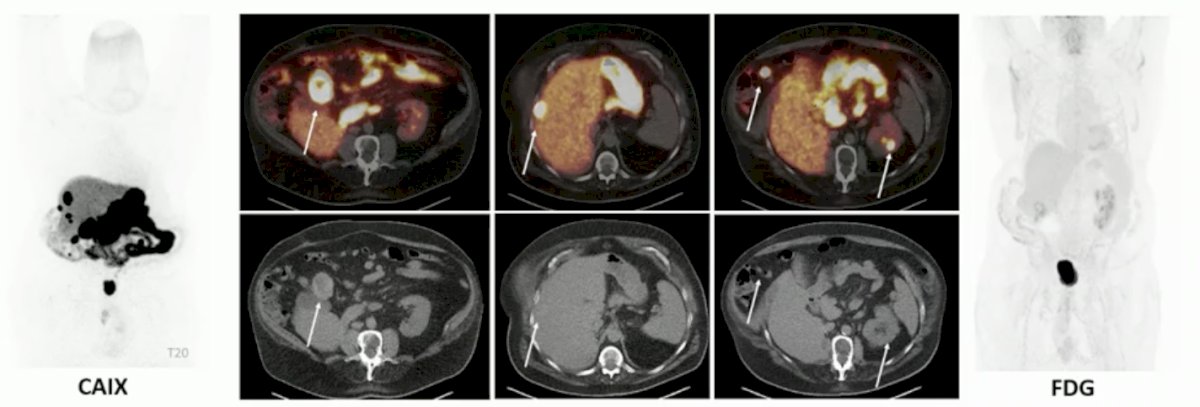
Dr. Küper highlighted growing enthusiasm for the [⁶⁸Ga]-CAIX radiotracer, given its superior ability to detect ccRCC lesions missed by conventional imaging. In one illustrative case (Figure below), a 61-year-old patient with previously resected ccRCC developed a mesenteric metastasis and a suspected liver lesion. While FDG-PET/CT failed to identify additional disease, [⁶⁸Ga]-CAIX PET/CT revealed multiple metastatic sites, shifting management from a planned local surgical approach to systemic therapy.
This case exemplifies how CAIX-targeted imaging can uncover occult disease, refine staging, and directly alter treatment decisions in advanced ccRCC.
Dr. Küper concluded her presentation by emphasizing that there remains a clear need for improved diagnostic and therapeutic strategies in ccRCC. [⁶⁸Ga]-CAIX PET/CT has emerged as a promising imaging tool, demonstrating superior lesion detection compared to CT, substantial clinical impact, and high SUV values that support its theranostic potential. Notably, 56% of patients exhibited sufficient CAIX expression to qualify for [¹⁷⁷Lu]-CAIX therapy under institutional criteria, with the first successful treatments already reported by Hofman et al. (1) in the ongoing ITM-sponsored phase I/II trial (NCT05706129).
Presented by: Alina T. Küper, MD, Resident, Department of Nuclear Medicine bei Universitätsklinikum Essen. Essen, Germany.
Written by: Julian Chavarriaga, MD – Urologic Oncologist at Cancer Treatment and Research Center (CTIC) via Society of Urologic Oncology (SUO) Fellow at The University of Toronto. @chavarriagaj on Twitter during the 2025 European Society for Medical Oncology (ESMO) Annual Congress, Berlin, Germany, October 17–21, 2025