(UroToday.com) The 2025 European Association of Urology (EAU) Annual Meeting held in Madrid, Spain was host to the session Progress and Controversies in oncological urology from the EAU Section of Oncological Urology. Dr. Daniela Oprea-Lager discussed the role of molecular imaging in the pre-treatment work-up of muscle invasive bladder cancer (MIBC).
Role of molecular imaging in MIBC
Dr. Oprea-Lager began her presentation by highlighting the multiple potential roles of molecular imaging in muscle-invasive bladder cancer (MIBC). These include primary tumor assessment (T stage), evaluation of lymph node metastases (N stage), and detection of distant metastases (M stage). Additionally, molecular imaging plays a crucial role in patient management at initial diagnosis, assessing recurrence after surgery, and evaluating response to neoadjuvant or induction chemotherapy.
Despite its potential, several challenges remain for molecular imaging in MIBC. Key needs include imaging modalities that integrate both anatomical and functional assessment, the development of hybrid techniques, sufficient signal strength for accurate detection, and suitable tracers specific to MIBC. Additionally, comprehensive imaging should allow for the detection of all metastatic sites and enable whole-body scanning to optimize disease evaluation and management.
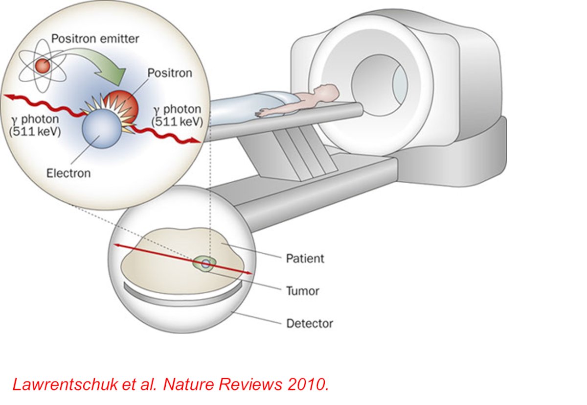
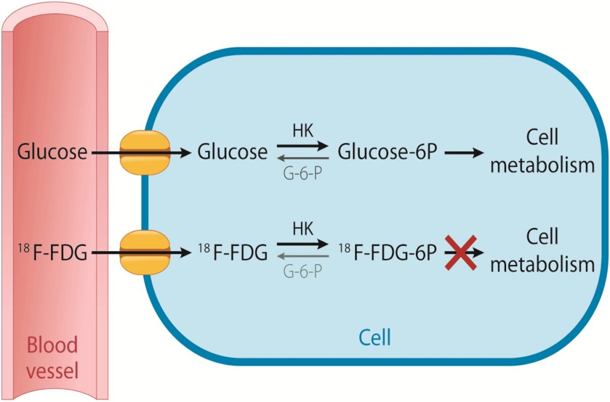
¹⁸F-Fluorodeoxyglucose (FDG) is a radiotracer used in positron emission tomography (PET) imaging that exploits the increased glucose metabolism characteristic of many cancer cells. Once administered, FDG accumulates in tissues with high metabolic activity, allowing for the visualization of both normal physiological processes and pathological conditions. In a standard FDG-PET scan, pronounced physiological uptake is observed in the brain and heart due to their high glucose consumption, with lesser uptake in the salivary glands. Additionally, because FDG is excreted through the urinary tract, notable tracer accumulation occurs in the bladder.
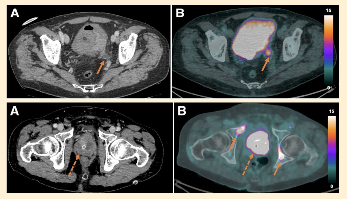
¹⁸F-fluorodeoxyglucose (¹⁸F-FDG) PET-CT has gained prominence in staging muscle-invasive bladder cancer (MIBC), particularly when conventional imaging yields inconclusive results. This modality is increasingly used to assess lymph node involvement and detect recurrence post-treatment. PET-CT enhances the characterization of abnormal lymph nodes and bony lesions, providing valuable diagnostic insights that may influence clinical decision-making, as shown below.
However, the question of whether ¹⁸F-FDG PET-CT is ready for prime time was summarized by Dr Oprea-Lager. She mentioned that in summary for TNM staging ¹⁸F-FDG PET-CT:
- T: has no established role in evaluation of a primary urothelial tumor.
- N: Shows an improvement in sensitivity for detecting pelvic lymph node involvement in bladder cancer compared to CT alone, Sensitivity of 56% vs 40%, respectively.
- M: Has shown superior performance in detection of distant metastases in urothelial carcinoma compared to CT/MRI. However, its use is not currently supported by guidelines.
The ideal alternative tracer for imaging in MIBC should possess a suitable structure for radiolabeling, stable labeling, and a well-defined molecular target. It should demonstrate high specificity for cancer biomarkers such as FAP, Nectin-4, CAIX, and uPAR, ensuring precise tumor detection. Additionally, the tracer should exhibit rapid urothelial tumor uptake, efficient clearance from the blood, an optimal half-life, and reduced urinary excretion to minimize background noise and enhance imaging quality.
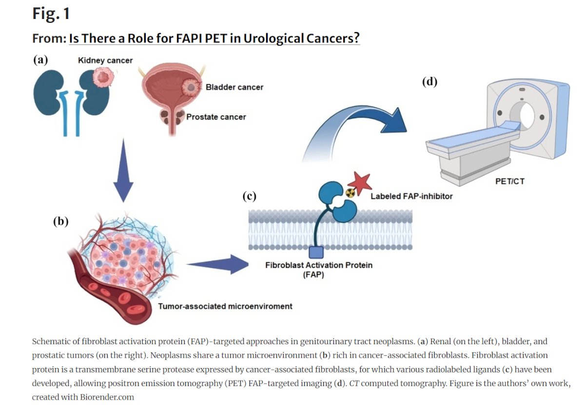
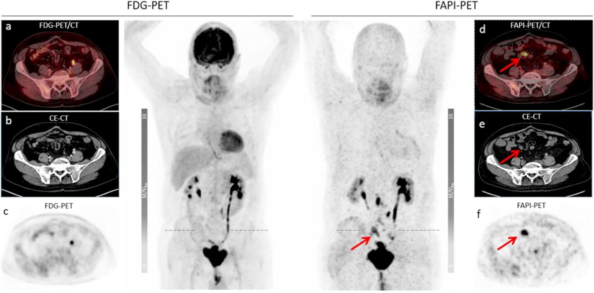
68Ga/18F-FAPI PETTumors share a unique microenvironment characterized by the presence of cancer-associated fibroblasts (CAFs), which play a key role in tumor progression. Fibroblast activation protein (FAP), a transmembrane serine protease, is highly expressed by CAFs but minimally present in healthy tissues. This makes FAP an attractive target for imaging and therapy. The ⁶⁸Ga/¹⁸F-FAPI PET tracer rapidly accumulates in lesions while maintaining low background activity, leading to a high tumor-to-background ratio and improved lesion detection. Given its specificity and potential for theranostic applications, FAPI PET is emerging as a promising tool for MIBC diagnosis and treatment planning.
This comparison highlights the potential of ⁶⁸Ga-FAPI PET/CT in bladder cancer imaging, particularly in detecting metastatic lesions that may not be visible on ¹⁸F-FDG PET/CT. The example of a 68-year-old patient undergoing follow-up after radical cystectomy illustrates the superior sensitivity of ⁶⁸Ga-FAPI in identifying mesenteric and abdominal lymph node metastases. These findings suggest that ⁶⁸Ga-FAPI PET/CT could offer improved accuracy in staging and restaging bladder cancer, potentially influencing treatment decisions.
CAIX is a highly promising biomarker in bladder cancer, given its expression in 70% to 90% of tumors while being absent in normal urothelial tissue. This selective expression makes it a valuable target for both diagnostic and therapeutic advancements. Radiolabeled girentuximab, an anti-CAIX antibody, enables in-vivo assessment of CAIX expression via PET imaging, offering a non-invasive method to better characterize urothelial carcinoma and potentially guide targeted treatment strategies.
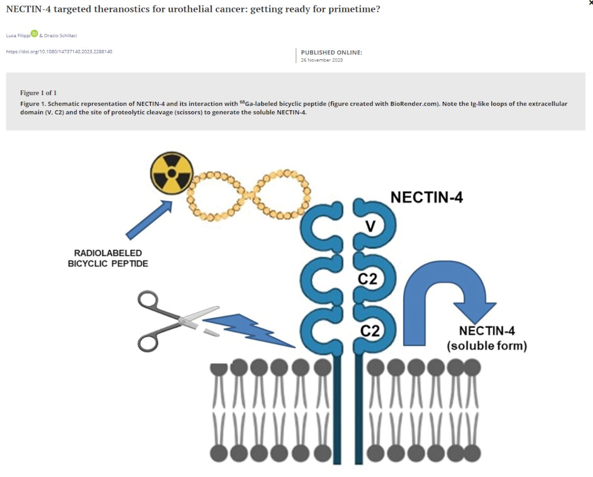
Nectine-4Nectin-4 is a tumor-associated antigen that has emerged as a promising target for molecular imaging in muscle-invasive bladder cancer (MIBC). Its extracellular region consists of three immunoglobulin (Ig) domains of both IgV and IgC types, and its soluble form is generated through metalloproteinase-mediated cleavage at the cell surface. These structural characteristics make Nectin-4 particularly well-suited for molecularly targeted imaging and therapeutic strategies. Notably, it is selectively overexpressed in bladder cancer, with approximately 83% of bladder tumors and up to two-thirds of upper tract urothelial carcinomas (UTUC) exhibiting Nectin-4 expression. Its strong association with poor prognosis further underscores its potential as a biomarker for both diagnostic and therapeutic applications.
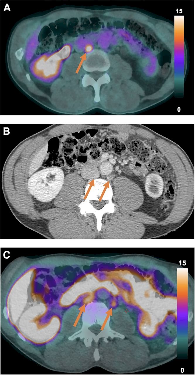
Copper plays a key role in cellular turnover, mitochondrial respiration, carcinogenesis, and cancer metabolism. It is essential for cytochrome-c oxidase activity and is a key factor in many tumoral pathways, including those in bladder cancer. The advantage of this tracer over others is its biodistribution, which is well-suited for pelvic molecular imaging, as ⁶⁴CuCl₂ is neither excreted nor accumulated in the urinary tract or bladder.
Dr. Oprea-Lager presented an example of a patient who underwent both ¹⁸F-FDG PET/CT and ⁶⁴CuCl₂ PET/CT. Notably, in the images below, ¹⁸F-FDG PET/CT (A) and ⁶⁴CuCl₂ PET/CT (C) revealed that the paracaval node showed uptake of both tracers, whereas the paraaortic node accumulated ⁶⁴CuCl₂ but not ¹⁸F-FDG (arrows), showing its potential role in improving MIBC staging.
Dr. Oprea-Lager wrapped up her presentation with the following conclusions:
- Molecular imaging is a rapidly evolving field in MIBC
- It offers improved diagnostic and prognostic capabilities
- 18F-FDG PET-CT has shown utility in staging
- Further prospective research is needed to validate new promising tracers such as FAPI, Nectine-4, CAIX, 64CuCL2
- Advances in theranostics and molecular imaging have the potential to revolutionize MIBC management, enhancing the ability to tailor treatments and improve patient outcomes
Presented by: Daniela-Elena Oprea-Lager, MD, PhD, Professor of Nuclear medicine at the Nijmegen University Medical Center, Dept of Medical Imaging. Nijmegen, Netherlands
Written by: Julian Chavarriaga, MD – Urologic Oncologist at Cancer Treatment and Research Center (CTIC) via Society of Urologic Oncology (SUO) Fellow at The University of Toronto. @chavarriagaj on Twitter during the European Association of Urology (EAU) 2025 Annual Meeting, Madrid, Spain, Fri, Mar 21 – Mon, Mar 24, 2025.