(UroToday.com) At the 38th Annual Meeting of the Engineering & Urology Society (EUS), Mr. Dan Luca of UCLA presented a compelling advancement in the imaging of prostate cancer (PCa), identifying prostate ductal anatomy as a novel contrast mechanism for micro-ultrasound (MicroUS). His team’s research proposes a method to enhance cancer detection in the prostate by visualizing structural disruptions in ductal architecture, features that are invisible with conventional ultrasound.
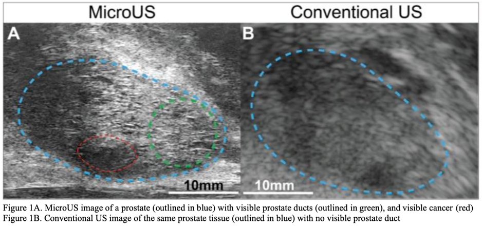
Conventional ultrasound, operating at 6-9 MHz with a resolution of roughly 200 µm, has long been criticized for its poor sensitivity and specificity in detecting clinically significant PCa.1,2 In contrast, MicroUS, with a significantly higher resolution of 70 µm at 29 MHz, enables real-time visualization of finer structures within the prostate, including ductal anatomy (Figure 1). This approach also has similar cancer detection rates to MRI. As such, this technology capitalizes on the fact that prostate cancer often disrupts the normal ductal network.2,3
In this IRB-approved pilot study, ten men scheduled for radical prostatectomy were enrolled. After surgery, their prostates were sectioned, stained, and analyzed with a custom MATLAB computer vision algorithm (Figure 2).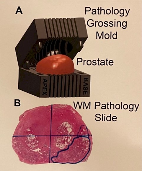
Figure 2A: Excised Prostate Grossed into Sections Using Grossing Mold
Figure 2B: Resulting WM Pathology Slide Annotated with Pca Region
The study evaluated 54 whole-mount pathology slides, which were scanned and annotated to distinguish benign tissue from cancerous lesions. A binary masking and color thresholding pipeline was used to quantify key features of the ducts, specifically the duct equivalent diameter and the ductal ratio (the proportion of white space representing ducts within the tissue) (Figure 3).
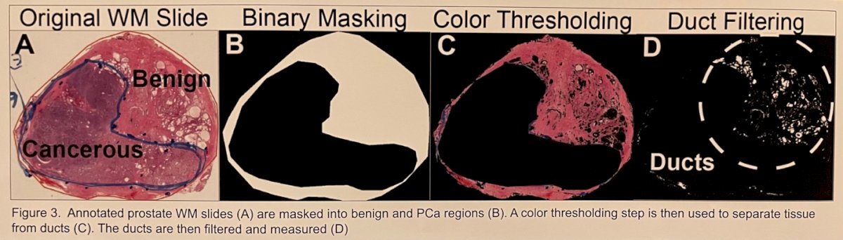
Figure 3: Annotated prostate WM slides (A) are masked into benign and PCa regions (B). A color thresholding step is then used to separate tissue from ducts (C). The ducts are then filtered and measured (D).
The team identified a total of 145,150 benign ducts and 9,245 cancerous ducts. Results showed that benign ducts had a significantly larger mean diameter (155.7 ± 147.8 µm) compared to cancerous ducts (129.4 ± 94.3 µm). To this end, while the mean diameter for both tissue types remains below the conventional ultrasound resolution threshold (200 µm), the MicroUS resolution threshold is lower than the mean diameters, therefore being easily detected with the latter method (Figure 4A). Additionally, the ductal ratio was more than halved in PCa tissue (0.039 ± 0.017) compared to benign areas (0.086 ± 0.029), with both findings achieving high statistical significance (p << 0.001) (Figure 4B).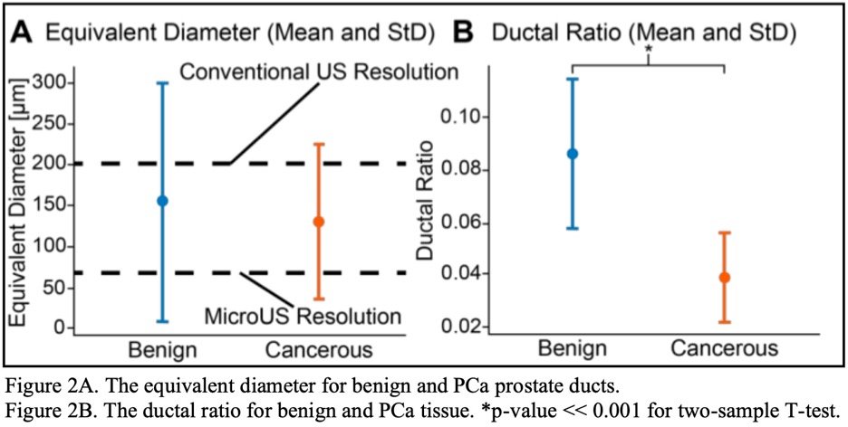
Figure 4A: The Equivalent Diameter for Benign and PCa Prostate Ducts.
Figure 4B: The ductal ratio for Benign and PCA Tissue. *P-value <<0.001 for two-sample T-test.
These findings suggest that disruptions in ductal anatomy, specifically a reduction in ductal density and size, may serve as a visual biomarker for cancerous regions on MicroUS, offering a highly promising contrast mechanism for PCa detection.
To close, Mr. Luca stated that conventional ultrasound fails to view a lot of these depths because it is too small, while MicroUS can visualize ductal anatomy as a contrast mechanism for prostate cancer visualization. This study offers a promising glimpse into a future where real-time, resolution-enhanced ultrasound could rival the diagnostic capabilities of more expensive or less accessible imaging modalities.
Moderator Dr. Rha asked Mr. Luca if future steps of this project include the augmentation of MRI scans, to which he responded that his work will propose MicroUS as a safe and cost-effective alternative to MRI.
Presented by: Dan Luca, University of California, Los Angeles
Co-Authors: Jake W. Pensa, Derrick Ushko, Griffith Hughes, Adam Kinnaird, David Kuppermann, Anthony Sisk, Leonard Marks, Rory Geoghegan, and Wayne G. Brisbane
Moderated by: Koon Ho Rha and Andreas Forsvall
Written by: Seyed Amiryaghoub M. Lavasani, B.A., University of California, Irvine, @amirlavasani_ on Twitter during the American Urological Association's 2025 Annual Meeting, between April 26 – 29, 2025, in Las Vegas, NV.
References:
- Hricak H, Choyke PL, Eberhardt SC, Leibel SA, Scardino PT. Imaging Prostate Cancer: A Multidisciplinary Perspective. Radiology. 2007;243(1):28-53. doi:10.1148/radiol.2431030580
- Klotz L, Lughezzani G, Maffei D, et al. Comparison of micro-ultrasound and multiparametric magnetic resonance imaging for prostate cancer: A multicenter, prospective analysis. Can Urol Assoc J. 2021;15(1): E11-E16. doi:10.5489/cuaj.6712
- Ghai S, Eure G, Fradet V, et al. Assessing Cancer Risk on Novel 29 MHz Micro-Ultrasound Images of the Prostate: Creation of the Micro-Ultrasound Protocol for Prostate Risk Identification. J Urol. 2016;196(2):562-569. doi:10.1016/j.juro.2015.12.093