(UroToday.com) On the first day of the 2024 American Urological Association (AUA) Annual Meeting, Loma Linda medical student Ruben Crew and his colleagues unveiled a pioneering approach integrating deep learning algorithm software and CT imaging for the detection of ureteral stones in the presence of metal hardware. Their study aimed to employ and compare deep learning reconstruction (DLR) and metal artifact reduction (MAR) software in assessing the quality of low dose and conventional CT scan imaging for ureteral stone detection in the presence of metal hip prostheses. Using a cadaver model with bilateral hip prostheses and ten stones implanted in the ureter near the ureterovesical junction via laparotomy, both conventional and ultra-low dose CT scans with and without DLR and MAR were performed. Two-blinded radiologists evaluated each set of scans for stone count and location. Objective and subjective comparisons were conducted using ROC curve analysis and a 5-point modified Likert scale, respectively. The author summarized the study's methodology in the following flow chart:

Figure 1. Summary of the method. UVJ= Ureterovesical Junction, CD= Conventional Dose, ULD= Ultra-Low Dose, DLR= Deep Learning Reconstruction, MAR= Metal Artifact Reduction (Crew et al.).
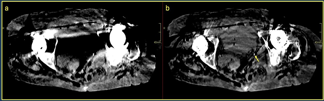
Figure 2. (A) Axial view of ULD alone. (B) Axial view of ULD with DLR and MAR with arrow pointing at the stone burden within the ureter (Crew et al.).
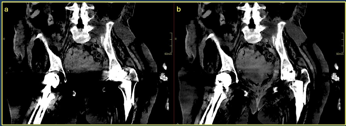
Figure 3. (A) Coronal view of ULD alone. (B) Coronal view of ULD with DLR and MAR (Crew et al.).
As depicted in Figures 2 and 3, the application of DLR and MAR notably improves the detection of ureteral stones in the presence of metal artifacts. Comparing ULD, ULD with DLR and MAR, CD, and CD with DLR and MAR, ROC analysis revealed significant differences. The incorporation of DLR and MAR into ULD CT not only significantly enhances stone detection accuracy (p<0.05, Figure 4), aligning it with CD and CD with DLR and MAR (p>0.25, Figure 4), but also elevates all image quality parameters (p<0.05, Figure 5), surpassing those of CD (p<0.05, Figure 5). In conclusion, this innovative investigation by Crew and his colleagues demonstrates that the utilization of DLR and MAR significantly reduces radiation exposure without compromising the accuracy of stone burden detection in patients with metal artifacts.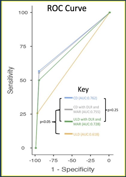
Figure 4. ROC Curve of Stone Detection in ULD, ULD with DLR and MAR, CD, and CD with ULD and DLR (Crew at al.).
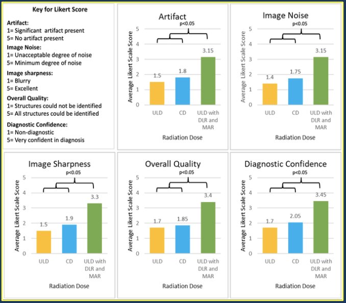
Figure 5. Comparison of average Likert Scale Score subjective parameters for ULD, ULD with DLR and MAR, CD, and CD with ULD and DLR (Crew et al.).
During the question-and-answer period, various aspects were explored regarding the study's methodology and implications. Firstly, concerns were raised about the absence of hydronephrosis or secondary symptoms in the cadaver model and its potential impact on algorithm sensitivity. However, Crew explained that the study's focused examination of the distal ureter minimized the likelihood of significant interference from hydronephrosis. Further inquiries centered on the location of stone placement and whether the radiologists were blinded to the location of placement. The presenter confirmed that radiologists were indeed blinded, and stones were strategically placed at the ureterovesical junction (UVJ), 2 cm and 4 cm above UVJ. Discussions also touched upon the deep learning algorithm specifics and if the scanning protocols would be different than regular CT-scan protocol. Crew responded that the CT-scan machine manufacturer provides the program, and it can be applied for post-processing scans. Additionally, the moderator asked whether patients with bilateral hip prostheses were mostly part of the elderly population and if there was a significant risk posed by conventional radiation that could lead to negative outcomes. Crew responded that despite age, there is a minimal risk of radiation-induced cancer, with studies also shedding light on the paradigm shift towards hip replacements in younger generations. Lastly, a question was asked on the rationale behind conducting the study on cadavers instead of patients, despite challenges in stone implantation and the methodology of the study. An answer was given that the investigators wanted to minimize radiation exposure (avoid any patient exposure) as part of the study required CT scanning.
Presented by: Ruben Crew, Medical Student at Loma Linda University Health, Loma Linda, CA
Written by: Seyedamirvala Saadat, B.S., Assistant Research Specialist, Department of Urology, University of California Irvine, @Val_Saadat on X during the 2024 American Urological Association (AUA) Annual Meeting, San Antonio, TX, Fri, May 3 – Mon, May 6, 2024.References:
- Brenner DJ, Hall EJ. Computed tomography--an increasing source of radiation exposure. N Engl J Med. 2007 Nov 29;357(22):2277-84. doi: 10.1056/NEJMra072149. PMID: 18046031.
- McLeavy CM, Chunara MH, Gravell RJ, Rauf A, Cushnie A, Staley Talbot C, Hawkins RM. The future of CT: deep learning reconstruction. Clin Radiol. 2021 Jun;76(6):407-415. doi: 10.1016/j.crad.2021.01.010. Epub 2021 Feb 23. PMID: 33637310.
- Mizuki M, Yasaka K, Miyo R, Ohtake Y, Hamada A, Hosoi R, Abe O. Deep Learning Reconstruction Plus Single-Energy Metal Artifact Reduction for Supra Hyoid Neck CT in Patients With Dental Metals. Can Assoc Radiol J. 2024 Feb;75(1):74-81. doi: 10.1177/08465371231182904. Epub 2023 Jun 30. PMID: 37387607.
- Hosoi R, Yasaka K, Mizuki M, Yamaguchi H, Miyo R, Hamada A, Abe O. Deep learning reconstruction with single-energy metal artifact reduction in pelvic computed tomography for patients with metal hip prostheses. Jpn J Radiol. 2023 Aug;41(8):863-871. doi: 10.1007/s11604-023-01402-5. Epub 2023 Mar 2. PMID: 36862290; PMCID:PMC10366278.
- Khan SR, Pearle MS, Robertson WG, Gambaro G, Canales BK, Doizi S, Traxer O, Tiselius HG. Kidney stones. Nat Rev Dis Primers. 2016 Feb 25;2:16008. doi: 10.1038/nrdp.2016.8. PMID: 27188687; PMCID: PMC5685519
- Negm AM, Beaupre LA, Goplen CM, Weeks C, Jones CA. A Scoping Review of Total Hip Arthroplasty Survival and Reoperation Rates in Patients of 55 Years or Younger: Health Services Implications for Revision Surgeries. Arthroplast Today. 2022 Jul 19;16:247-258.e6. doi: 10.1016/j.artd.2022.05.012. PMID: 36092132; PMCID:PMC9458900.
- Shichman I, Roof M, Askew N, Nherera L, Rozell JC, Seyler TM, Schwarzkopf R. Projections and Epidemiology of Primary Hip and Knee Arthroplasty in Medicare Patients to 2040-2060. JB JS Open Access. 2023 Feb 28;8(1):e22.00112. doi: 10.2106/JBJS.OA.22.00112. PMID: 36864906; PMCID: PMC9974080.