(UroToday.com) The 2025 ASTRO annual meeting featured a prostate cancer session and a presentation by Dr. Jasmesh Sandhu discussing a comparison of PSMA PET/CT and MRI in prostate radiotherapy planning. In prostate radiotherapy, accurately identifying seminal vesicle involvement, or T3b disease, is critical for effective treatment planning. Seminal vesicle invasion indicates more advanced prostate cancer and may necessitate a change in radiotherapy target volume. Importantly, missing seminal vesicle invasion can lead to under-treatment of the cancer and an increased likelihood of future cancer recurrence. Multiparametric magnetic resonance imaging (mpMRI) is recommended as the standard imaging modality pre-biopsy in patients with a high PSA suspicious of prostate cancer and is regarded as the most accurate for T-staging. Recently, PSMA PET/CT has shown promising results as a tool for staging high-risk prostate cancer. The question remains as to whether one modality is better than the other at predicting seminal vesicle involvement. The aim of this study, presented at ASTRO 2025, was to compare the accuracy and reliability of PSMA PET/CT and mpMRI in detecting seminal vesicle involvement to optimize prostate radiotherapy planning.
A multicenter, international, retrospective study was conducted at Guy’s and St Thomas’ Hospital, London, and San Raffaele Institute, Milan. Men who underwent radical prostatectomy for biopsy proven prostate cancer between 2018 and 2023 were identified. Collected data included histopathological findings, such as T3b disease, Gleason score, PSA levels, margin positivity, and pre-operative mpMRI and 68Ga-PSMA-11 PET/CT imaging. Whole-mount pathology served as the reference standard. Data analysis was performed in London using statistical software.
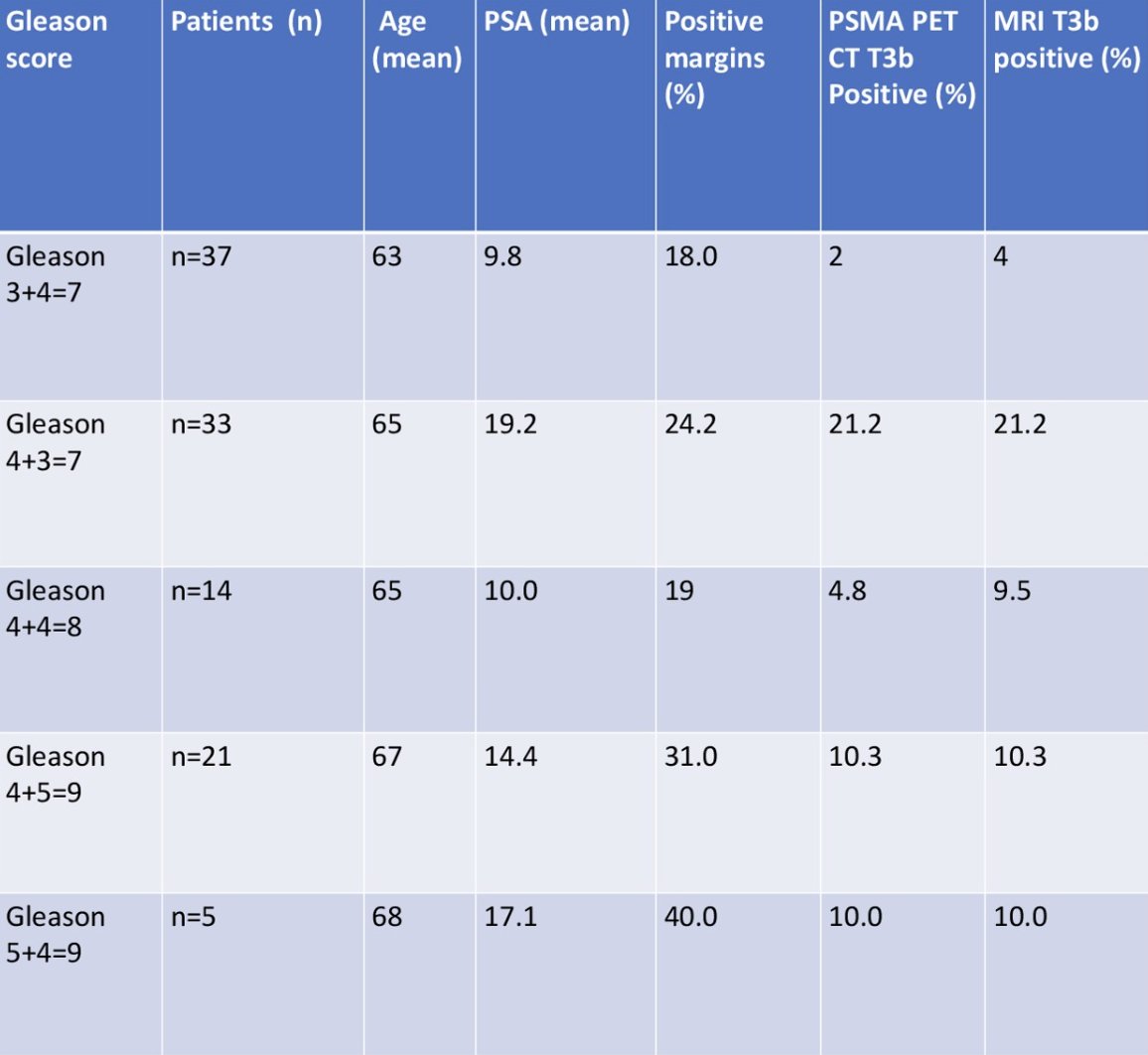
Among 268 men (median age 65.4 ± 1.4 years), 110 underwent both PSMA PET/CT and mpMRI. Histologically verified seminal vesicle involvement was found equally in 17.3% of patients who underwent PSMA PET/CT or mpMRI. Sensitivity was 16.7% (95% CI 10.87%–23.95%) for PSMA PET/CT and 13.7% (95% CI 9.32%–19.22%) for mpMRI. Specificity was 100% for both modalities. Positive predictive value was 100% for both modalities, and negative predictive value was 37.8% (PSMA PET/CT) versus 28.5% (mpMRI). The area under the ROC curve (AUC) was 0.586 (PSMA PET/CT) versus 0.606 (mpMRI). Moreover, there was no statistically significant difference in diagnostic accuracy between the 2 modalities. Multivariate analysis showed no significant correlation between age, PSA, and T3b disease (p = 0.420). The following table represents a subgroup analysis exploring the correlation between histologically confirmed T3b disease on PSMA PET/CT and MRI with age, margin positivity, Gleason score, and PSA:
Dr. Sandhu concluded this presentation discussing a comparison of PSMA PET/CT and MRI in prostate radiotherapy planning with the following take home points:
- No significant difference was observed between mpMRI and PSMA PET/CT in detecting T3b disease
- The key limitation of this study was a small sample size
- Further studies are required to refine imaging strategies for prostate radiotherapy planning
Presented by: Jasmesh Sandhu, MB, ChB, NHS London, London, UK
Written by: Zachary Klaassen, MD, MSc – Urologic Oncologist, Associate Professor of Urology, Georgia Cancer Center, Wellstar MCG Health, @zklaassen_md on Twitter during the 2025 American Society for Radiation Oncology (ASTRO) Annual Meeting, San Francisco, CA, September 28th – 30th, 2025