(UroToday.com) The 2025 American Society of Clinical Oncology Genitourinary (ASCO GU) cancers symposium held in San Francisco, CA was host to the Poster Session A: Prostate Cancer. Dr. Aidan McLoughlin presented Abstract 335: Comparison of digital pathology AI models, genomics classifier, and clinical variables in predicting progression free survival in TCGA prostate data set.
Clinical pathological risk factors (such as Gleason score, seminal vesicle invasion pT3b, and extraprostatic extension), genomic classifiers (GC), and recently developed digital pathology AI (DPAI) models have been used to predict disease progression or survival outcomes. The investigators aimed to address key gaps in DPAI models applied to survival analysis using hematoxylin and eosin whole slide images from The Cancer Genome Atlas (TCGA) prostate dataset. While some AI models have been developed for survival analysis with TCGA data, there is limited understanding of how these models predict progression-free survival (PFS) in prostate cancer. Additionally, previous studies have not comprehensively compared DPAI models to genomic or clinical models, nor have they accounted for batch effects. This study directly compared DPAI models, genomic classifiers, and clinical variables in predicting PFS in the TCGA prostate dataset.
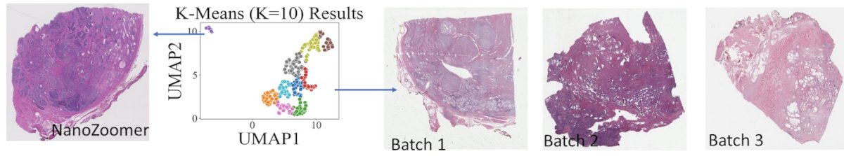
The TCGA prostate dataset (PRAD) includes 390 patients with whole slide images, gene expression, and clinical data (pT3b, Gleason, EP, N+), along with time from surgery to assess PFS. To evaluate potential batch effects in the whole slide images, the investigators used HistoQC. They applied linear Cox regression for clinical variables and utilized a 5-gene model from Mou et al.1 as an example genomic classifier.
DPAI model components were:
- Image encoders: ResNet50 based on Image Net (IN), UNI
- WSI models: global average, CLAM, PatchGCN, HVTSurv.

They then trained four different digital pathology AI models to predict progression-free survival using five-fold cross-validation, ensuring that batch effects were accounted for in the data splits. This cross-validation process was repeated five times with different random splits. Each model’s performance was assessed using the mean concordance index (C-index), with confidence intervals approximated by taking the second smallest to the second largest value from the 25 splits.
Additionally, they evaluated multi-modal models that combined clinical variables with gene expression and/or digital pathology AI features, following the same performance assessment methodology.
The investigators observed substantial whole slide image batch effects across the PRAD dataset introduced by a scanner (NanoZoomer vs Aperio), leading to the exclusion of nine patients.
Among the remaining 383 patients, the clinical-only model achieved a mean C-index of 0.73 (0.63–0.84) in cross-validation, while the expression-only model (Genomic) performed similarly at 0.72 (0.58–0.88). However, the combined clinical-genomic model demonstrated superior performance, achieving a C-index of 0.79 (0.65–0.90).

Among the digital pathology AI models, the clustering-constrained-attention multiple-instance learning (CLAM) model—an attention-based approach that identifies diagnostically relevant subregions within whole slide images and refines the feature space through instance-level clustering—achieved the highest mean C-index of 0.68 (0.57–0.82) when initialized under the UNI foundation model in this study.3 Combining this model with clinical variables modestly improved performance to a C-index of 0.71 (0.59–0.83). Notably, adding gene expression data to this multi-modal model did not provide additional benefit.

Dr. McLoughlin concluded their poster with the following key takeaways:
- This study represents the first in-depth analysis of PFS prediction in PRAD, comparing DPAI models with clinical and gene expression-based models.
- Different modalities of data (clinical, genomic, digital pathology) achieve similar performances in TCGA prostate dataset, while the combination clinical-genomic model has the best performance in predicting PFS
- All data modalities demonstrated similar C-indices with overlapping confidence intervals, though the clinical-genomic model performed best.
- Controlling for batch effects is crucial for DPAI models. Future research should explore whether batch effect correction in WSI can improve DPAI performance to match clinical-genomic models.
- Hierarchical Al modeling approaches have similar performance. Pathology specific foundation model negates performance differences between naïve and complex Al models.
- A larger sample size is needed to adequately compare data modalities and develop robust multi-modal models.
Presented by: Aidan McLoughlin, Biostatistics PhD Student at University of California Berkeley, CA, United States.
Written by: Julian Chavarriaga, MD – Urologic Oncologist at Cancer Treatment and Research Center (CTIC) via Society of Urologic Oncology (SUO) Fellow at The University of Toronto. @chavarriagaj on Twitter during the 2025 Genitourinary (GU) American Society of Clinical Oncology (ASCO) Annual Meeting, San Francisco, CA, Thurs, Feb 13 – Sat, Feb 15, 2025.
References:- Mou Z, Spencer J, Knight B, John J, McCullagh P, McGrath JS, Harries LW. Gene expression analysis reveals a 5-gene signature for progression-free survival in prostate cancer. Front Oncol. 2022 Aug 12;12:914078. doi: 10.3389/fonc.2022.914078. PMID: 36033512; PMCID: PMC9413154.
- Janowczyk A, Zuo R, Gilmore H, Feldman M, Madabhushi A. HistoQC: An Open-Source Quality Control Tool for Digital Pathology Slides. JCO Clin Cancer Inform. 2019 Apr;3:1-7. doi: 10.1200/CCI.18.00157. PMID: 30990737; PMCID: PMC6552675.
- Lu MY, Williamson DFK, Chen TY, Chen RJ, Barbieri M, Mahmood F. Data-efficient and weakly supervised computational pathology on whole-slide images. Nat Biomed Eng. 2021 Jun;5(6):555-570. doi: 10.1038/s41551-020-00682-w. Epub 2021 Mar 1. PMID: 33649564; PMCID: PMC8711640.