

(UroToday.com) The 2025 American Society of Clinical Oncology (ASCO) Genitourinary (GU) Annual Symposium held in San Francisco, CA was host to a prostate cancer poster session. Dr. Jesus Casillas presented a study evaluating the association of PSMA PET results at biochemical recurrence (BCR) with metastasis free survival (MFS) by conventional imaging (CI) in patients with locally advanced or high-risk localized prostate cancer initially treated with radical prostatectomy.
Locally advanced, high-risk prostate cancer (LAHR PCa) comprises 10%-15% of new prostate cancer diagnoses in the U.S. and carries a higher risk of BCR, up to 60% following definitive treatment, compared to low-risk disease.1
Conventional imaging, including computed tomography (CT), magnetic resonance imaging (MRI), and single-photon bone scans, often fails to detect sites of disease recurrence at lower PSA levels during BCR.2 Prostate-specific membrane antigen (PSMA) positron emission tomography-computed tomography (PET-CT) is used to stage PCa at BCR and has a higher sensitivity than conventional imaging (i.e. CT and bone scan).3,4
There is a significant lack of evidence on how PSMA PET findings affect treatment decisions,3 including strategies such as radiation, androgen deprivation therapy, or systemic therapies and their timing; as well as the resulting clinical outcomes in patients with conventional imaging-defined LAHR PCa experiencing BCR after radical prostatectomy. To this end, Dr. Casillas and colleagues investigated the association between PSMA PET-CT results and metastasis-free survival (MFS) by conventional imaging in LAHR PCa patients with BCR who had undergone a radical prostatectomy.
Patients with LAHR PCa (characterized by either PSA >20 ng/mL, Gleason Score 8-10, or tumor staging T4a or higher on initial diagnosis and no evidence of metastasis) who experienced BCR following radical prostatectomy and underwent a PSMA PET-CT scan were retrospectively identified from two academic centers in the United States between January 2016 and January 2024. PSMA PET+ status was defined as having evidence of a distant lesion by PSMA PET.
Treatment changes were recorded from the time of BCR and up to 60 days post-BCR. MFS was estimated by conventional imaging (CT and bone scan). Time-to-event analysis comparisons were performed between patients with PSMA PET positive (PSMA PET+) and PSMA PET negative (PSMA PET-) lesions to estimate the effect of imaging results on MFS. A 1:1 propensity score matching was performed to control for confounding factors. The propensity score is defined as the probability of being assigned to PSMA PET+ group conditioning on the PSA and treatment change at BCR. This probability is estimated using logistic regression.
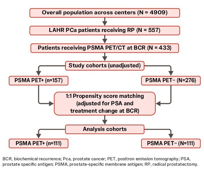
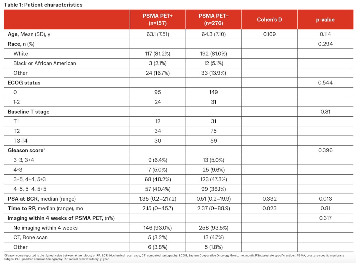
Four hundred thirty-three HR radical prostatectomy patients with mCRPC who had undergone a PSMA PET-CT at BCR were included. Of 433 patients, 157 were PSMA PET+.
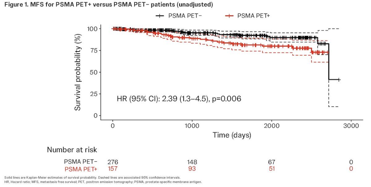
The overall median follow-up time was 47.3 months (interquartile range [IQR]: 21.2–72.8). MFS was significantly shorter for PSMA PET+ versus PSMA PET- patients by conventional imaging (p=0.006; HR: 2.39, 95% confidence interval [CI]: 1.3-4.5).
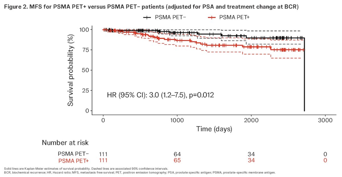
The difference in MFS remained significant after propensity score matching (p=0.012; HR: 3.0, 95% Cl: 1.2-7.5).
Dr. Casillas concluded as follows:
- Incorporating PSMA PET-CT results at time of BCR may enable more precise and effective treatment strategies
- Patients with locally advanced, high-risk PCa who have PSMA PET+ lesions at BCR following radical prostatectomy experience an MFS period three times shorter compared to patients without PSMA PET+ lesions at BCR
- A longer follow-up period is required to better evaluate associations with overall survival in this patient population
- Further analyses with a larger patient population across institutions in the United States and Europe are ongoing to increase the robustness of these MFS rate estimates
Presented by: Jesus Eduardo Juarez Casillas, MD, Resident Physician, Radiation Oncology, UCLA, Los Angeles, CA
Written by: Rashid K. Sayyid, MD, MSc – Robotic Urologic Oncology Fellow at The University of Southern California, @rksayyid on Twitter during the 2025 Genitourinary (GU) American Society of Clinical Oncology (ASCO) Annual Meeting, San Francisco, CA, Thurs, Feb 13 – Sat, Feb 15, 2025.
References:
- Shore ND, et al. Biochemical recurrence in patients with prostate cancer after primary definitive therapy: treatment based on risk stratification. Prostate Cancer Prostatic Dis. 2024; 27(2):192-201.
- Mena E, et al. Novel PET imaging methods for prostate cancer. World J Urol. 2021; 39(3):687-699.
- Meijer D, et al. Prostate-specific Membrane Antigen Positron Emission Tomography/Computed Tomography Is Associated with Improved Oncological Outcome in Men Treated with Salvage Radiation Therapy for Biochemically Recurrent Prostate Cancer. Eur Urol Oncol. 2022; 5(2):146-52.
- Hoffman A, et al. The Impact of PSMA PET/CT on Modern Prostate Cancer Management and Decision Making—The Urological Perspective. Cancers (Basel). 2023; 29;15(13):3402.