(UroToday.com) The 2025 American Society of Clinical Oncology (ASCO) Genitourinary (GU) Annual Symposium held in San Francisco, CA was host to a prostate cancer poster session. Dr. Isadora Martins de Sousa presented a study evaluating the prognostic utility of conventional imaging-defined volume and risk criteria to PSMA PET imaging findings in metastatic castration-sensitive prostate cancer (mCSPC) patients.
Dr. de Sousa noted that the treatment of mCSPC has evolved over the past decade. These therapies are guided by current volume (i.e., CHAARTED) and risk (i.e., LATITUDE) criteria, instituted based on conventional imaging findings (i.e., magnetic resonance imaging [MRI], computed tomography [CT], and bone scintigraphy [BS]). The emergence of novel imaging modalities, such as PSMA PET, with its greater accuracy and ability to detect metastatic disease earlier, raises questions about the applicability of these criteria in this context for earlier initiation of systemic therapy and the ability to distinguish groups with different survival outcomes.
To this end, the study investigators conducted a retrospective, single-center study including patients staged with PSMA PET between August 2017 and February 2022 and classified as having metastatic disease – either de novo (i.e., synchronous metastases) or recurrent (i.e., metachronous). Patients with node-only metastases were included. Patients were stratified into high and low volume and risk categories, using CHAARTED and LATITUDE classifications, respectively. The primary study outcome was overall survival.
This study included 48 patients with a median age of 66 years at diagnosis. 81.3% had Gleason scores between 8 to 10, 57% had ≥T3 disease, and 32% were cN1 by conventional exams. According to PSMA PET, 40% were de novo metastatic, with disease sites being bones (M1b - 52%), non-pelvic lymph nodes (Mla - 21%), pelvic lymph nodes (N1 - 17%), and viscera (M1c - 10%). 71% of them were categorized as low volume and 75% were low risk.
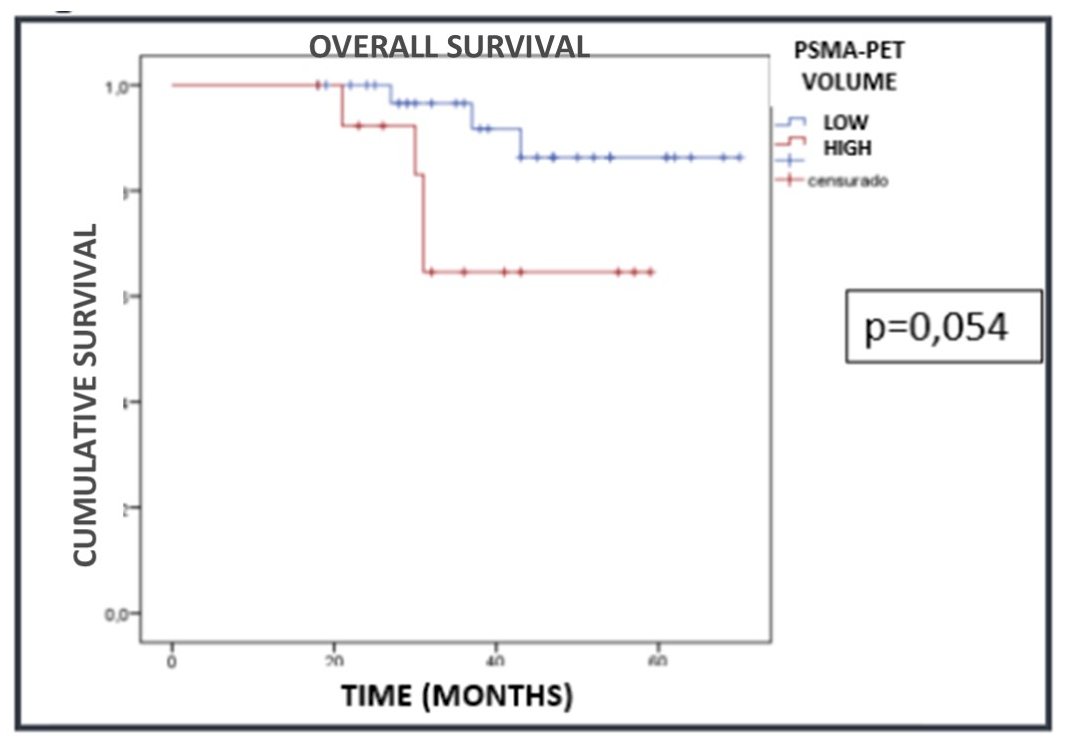
With a median follow-up of 37.5 months, with 7 deaths observed, the 36-months overall survival rates in the high- and low-volume populations were 64.6% and 96.6%, respectively (p=0.054).
The corresponding rates for low- and high-risk patients were 90% and 78%, respectively (p=0.74).
Overall, there was a 67% improved detection for distant metastatic disease with PSMA PET.

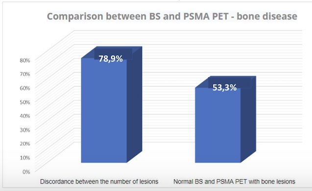
When correlated with bone scintigraphy, there was a 79% discordance between the number and/or location of bone lesions. In 53.3% of cases, PSMA PET was positive for bone metastases, despite concurrent negative bone scan findings.
Dr. de Sousa concluded that conventional imaging-defined volume criteria, but not risk criteria, are prognostic for overall survival when applied to PSMA PET findings in PET-staged metastatic prostate cancer patients. This study additionally confirms the superior diagnostic performance of PSMA PET, compared to conventional imaging, for detecting metastatic disease, including bone metastases.
Presented by: Isadora Martins de Sousa, AC, Camargo Cancer Center, São Paulo, Brazil
Written by: Rashid K. Sayyid, MD, MSc – Robotic Urologic Oncology Fellow at The University of Southern California, @rksayyid on Twitter during the 2025 Genitourinary (GU) American Society of Clinical Oncology (ASCO) Annual Meeting, San Francisco, CA, Thurs, Feb 13 – Sat, Feb 15, 2025.