(UroToday.com) In this presentation, Dr. W. Kimryn Rathmell first discussed various genetic pathways that have been identified in renal cell carcinoma (RCC) and related these pathways to ongoing work trying to utilize imaging technologies and other noninvasive strategies for improved RCC classification and detection.
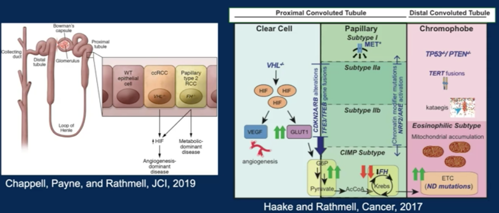
She first described the fundamental pathways leading to RCC development. For clear cell RCC, which arises in the proximal tubule, the canonical alterations occur on chromosome 3p. These affect the VHL gene, thus leading to upregulation of the hypoxia inducible factors and downstream signaling that leads to angiogenesis and other transcriptional outputs driving carcinogenesis and cell growth. She also described other molecular subtypes seen in RCC, including translocation driven RCC, fumarate hydratase mutation driven FCC, and alterations in PTEN and TP53 that are associated with chromophobe RCC that arises in the distal convoluted tubule.
Dr. Rathmell then described the first generation of tumor specific biomarkers that were published in 2014, which classified clear cell RCC (ccRCC) into group A (classical, angiogenic) and B (bad prognosis, upregulation of EMT), and these groups were prognostic for recurrence and disease-associated survival. She went on to describe how modern MRI imaging generates data beyond anatomic characterization of tumors that can be related to molecular characteristics of the tumor. In one study,1 features from MRI were identified (textural) that correlated with the group A and group B prognostic subtypes. More advanced MRI technologies such as modeling of hyperpolarized lactate signal and arterial spin labeling perfusion MRI are being analyzed for their ability to identify tumor characteristics that predict response to therapy. Similarly, PET tracers are being created to facilitate immunoPET (zirconium labeled atezolizumab) or exploit metabolic vulnerabilities such as glutamine addiction (radiolabeled glutamine) for more advanced imaging. Finally, Dr. Rathmell discussed the development and study of contrast enhanced ultrasound with infused microbubbles to identify more information about the contextual architecture of tumors.
The presentation then shifted to a discussion about other minimally invasive detection technologies. These include circulating tumor cells, cell free DNA, and cell-free methylated DNA immunoprecipitation.2
In the future, Dr. Rathmell envisions imaging or blood/urine-based assays being used to accurately detect RCC and potentially stratify tumors into treatment paths based on certain data characteristics.
Presented by: W. Kimryn Rathmell, MD, PhD, Director of the Division of Hematology and Oncology, Cornelius Abernathy Craig Chair, Professor of Medicine, Department of Clinical Medicine, Professor of Biochemistry, Professor of Cancer Biology, Vanderbuilt Unviersity Medical Center
Written by: Alok Tewari, MD, PhD, Medical Oncologist at the Dana-Farber Cancer Institute during the 2021 American Society of Clinical Oncology Genitourinary Cancers Symposium (#GU21), February 11th-February 13th, 2021
References:
1. Yin Q, Hung S-C , Rathmell W-K, et al., Integrative radiomics expression predicts molecular subtypes of primary clear cell renal cell carcinoma. Clin Radiol. 2018;73(9):782-791
2. Pier Vitale Nuzzo, Jacob E. Berchuck, Keegan Korthauer et al., Detection of renal cell carcinoma using plasma and urine cell-free DNA methylomes. Nature Medicine. 2020;26: 1041–1043