As a consequence, urinary toxicity after RP is common and may manifest in various forms, most notably urinary incontinence and urethral strictures. With the increasing use of stereotactic body radiotherapy (SBRT), dose-escalation, and reirradiation within the prostate bed (PB), standardization of the definition of urinary organs at risk (OARs) in the post-RP setting is needed.
According to GFRU experts, the delineation of urinary OARs is recommended in case of PB reirradiation with SBRT and PB SBRT. An agreement was reached to perform the delineation of urinary OARs in case of dose-escalated RT within the PB. The following structures were deemed as being critical to consider: bladder, bladder neck, bladder trigone, VUA, membranous urethra, and striated sphincter. Dose constraints were proposed and discussed for both reirradiation with SBRT and SBRT within the PB. Specifically, the role of the VUA in the development of toxicity following reirradiation with SBRT is discussed. As a tumor located in close proximity to the VUA has been shown to be associated with the onset of severe GU toxicity following reirradiation using SBRT, compromise could be made in the reirradiation setting to spare the VUA, even if it comes at the cost of a reduced target coverage. While dose constraints are implemented within prospective trials, their validity is, however, untested, and further studies assessing dosimetric predictors of toxicity following reirradiation and PB SBRT are awaited.
While several guidelines exist for the delineation of the PB in the postoperative setting, this consensus also discusses the optimal target volume for PB RT. Indeed, the new PERYTON guidelines proposed a new target volume based on recurrence patterns observed with PSMA PET/CT, which raises the possibility of sparing the bladder neck. Bladder neck sparing could be of interest in the postoperative setting. During RP, the lowest part of the bladder neck, including the internal urethral sphincter, is often resected, and therefore, the bladder neck’s height might rely on the preservation or reconstructive strategy used. (50). With these new guidelines, a large portion of the bladder neck (as well as the pubovesical and puboprostatic ligaments) is expected to be spared from radiation. The implementation of dose constraints allowing for a reduction of the bladder neck receiving the prescription dose could be meaningful, considering the role of the bladder neck for continence. Sparing the anterior portion of the bladder might be meaningful in terms of functional outcomes, especially in terms of anterior reconstruction. However, the hypothesis of a reduction in the rates of incontinence following PB RT with bladder-neck sparing is untested. Last but not least, sparing the bladder neck could be meaningful considering that this OAR is at risk of contracture following PB RT.
For the planning of routine postoperative RT, a CT scan without contrast agent is the recommended imaging modality. However, when delineation of urinary OARs is indicated, match-paired T2-weighted MRI is the recommended imaging associated imaging modality.
New clinical scenarios at risk of higher induced toxicity in the post-prostatectomy setting are arising, such as PB reirradiation with SBRT, PB SBRT, and dose-escalated RT within the PB. This consensus highlights contemporary urinary structures in the post-prostatectomy setting, to be considered at the time of treatment planning. It also provides a standardized definition of urinary OARs, which might be used in future clinical trials. While no validated dose constraints currently exist for urinary OARs - particularly the VUA, a frequent site of stricture and toxicity - these guidelines offer a foundation for future research. Integration of this consensus into clinical practice and prospective trials is essential to optimize therapeutic outcomes and minimize complications.
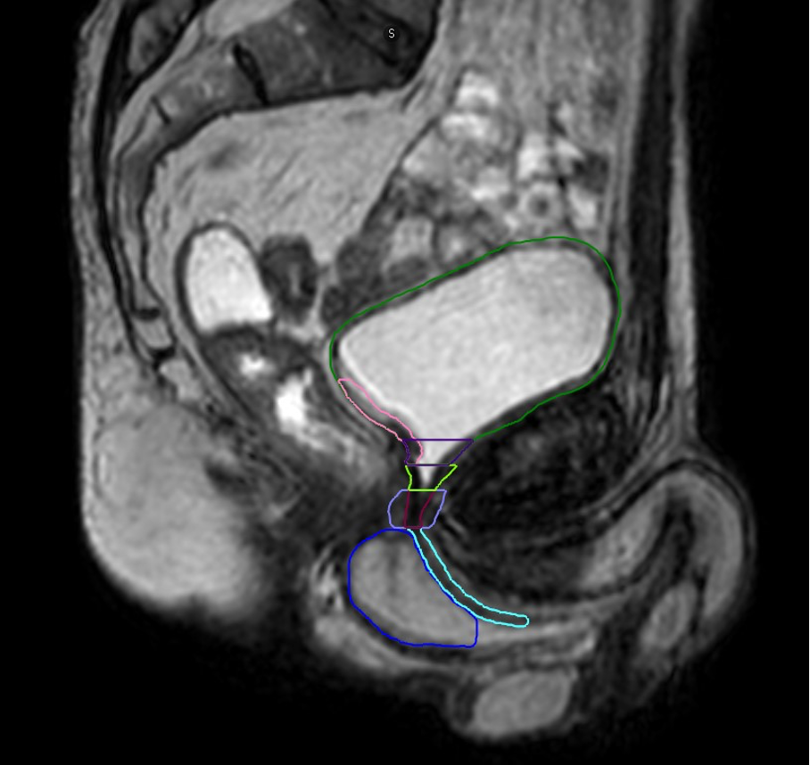
Figure 1: Illustration of urinary organs at risk following radical prostatectomy (T2-weighted sequence)

Figure 2: Illustration of the delineation of the prostate bed clinical target volume, according to the GFRU and PERYTON guidelines.
Dark green: bladder/ orange: ureters/ pink: bladder trigone/ purple: bladder neck/ light green: vesicourethral anastomosis/ burgundy: membranous urethra/ light purple: striated sphincter / light blue: bulbous urethra/ dark blue: penile bulb
In orange: prostate bed clinical target volume according to the PERYTON guideline. In pink: prostate bed clinical target volume according to the GFRU guideline.
Written by: Jennifer Le Guévelou, MD, PhD student, Laboratoire traitement du signal et de l'image, Université de Rennes, France
Read the Abstract