Within the miR-371-373 cluster, miR-371a-3p shows remarkable sensitivity and specificity for the detection of TGCT, overcoming current limitations of the classical serum tumor markers (HCG, AFP and LDH).3 The potential of miR-371a-3p has been demonstrated in several studies with multicentric retrospective and prospective patient cohorts, leading to its approval as an in vitro diagnostic (IVD) test for use in the clinic (already implemented and routinely reported at the Department of Pathology of IPO Porto for all patients with TGCTs).3,4 However, despite the established clinical utility, the secretion dynamics of these miRNAs in extracellular vesicles (EVs) derived from TGCT patients are still largely unknown. EVs are small lipid bound particles secreted by cells into the extracellular space, which can contain different types of cargo, from DNA to different types of RNAs, including microRNAs.5 The study of these small nanoparticles is important to understand tumor progression, since EVs are used by tumor cells for intercellular communication, for interacting with the tumor microenvironment, and for establishing pre-metastatic niches.6
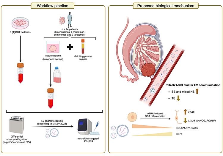
In this work, we successfully isolated and characterized EVs released from a wide range of TGCT samples (following MISEV guidelines), including cell lines, conditioned media from tissue explants (both tumor and adjacent testicular parenchyma), and matched plasma samples from the same patients, as well as age-matched male healthy blood donors. We then assessed microRNA expression (miR-371-373 cluster and let-7e), aiming to determine if these miRNAs are secreted in EVs derived from TGCT patient samples, and to verify if levels are concordant between matched tissue, blood sample sets, and their respective EV-secreted miRNAs.
In summary, our study showed that EVs from TGCT cell lines and clinical samples depict high levels of miR-371-373 cluster, mirroring their cellular quantities, with a clear shift in the EV-miRNA levels with differentiation, showing the absence of these miRNAs in teratoma. To our knowledge, this is the first study validating that TGCT-derived EVs from clinical samples (matched tissue explants and plasma) indeed carry high levels of miR-371-373 cluster, suggesting there may be an EV-based communication mechanism involving these miRNAs from the tissue to circulation.
Importantly, a microRNA switch is seen upon cell differentiation induced with ATRA, which triggers downregulation in the pluripotency-related miR-371-373 cluster, downregulation of LIN28 and other pluripotency-related markers, and upregulation of let-7e. Consequently, although biomarker performance suggests that EV-miR-371a-3p may not be the best candidate to use in the clinic when compared with total circulating miR-371a-3p, the observed changes in the miRNA cargo of EVs highlight their utility as indicators of TGCT cell state and mediators of intercellular communication and phenotype switch.
Furthermore, these EVs may be used in the future as therapeutic targets, as they are suggested to deliver oncogenic cargo to near and distant tumor microenvironment cells.5 The pipeline (including pre-clinical and matched tissue-plasma clinical samples) and consequent results herein presented could be used in the future for the discovery of novel clinical EV-derived biomarkers and to deepen our understanding of TGCT biology, namely in the emergence of teratoma.
This work was developed at the Cancer Biology and Epigenetics Group of the Research Center of IPO Porto and at the Department of Pathology of IPO Porto, in collaboration with the Urology Clinic and collaborators from the University of Cornell and California (USA).
Written by: Nuno Tiago Tavares,1,2 Catarina Lourenço,1-4 Vera Constâncio,1,2 Fernanda Fernandes-Pontes,1 Diana Fonseca,1 Rui Silva-Santos,5 Isaac Braga,6 Joaquina Maurício,7 Rui Henrique,1,5,8 Michelle Liu,9 Robert S Weiss,9 Aditya Bagrodia,10 Carmen Jerónimo,1,8 João Lobo1,5,8
- Cancer Biology and Epigenetics Group, Research Center of IPO Porto (CI-IPOP) / CI-IPOP@RISE Health Research Network - Portuguese Oncology Institute of Porto (IPO Porto) / Porto Comprehensive Cancer Center Raquel Seruca (Porto.CCC Raquel Seruca), Porto, Portugal.
- Doctoral Programme in Biomedical Sciences, School of Medicine and Biomedical Sciences, University of Porto (ICBAS-UP), Porto, Portugal.
- i3S - Instituto de Investigação e Inovação Em Saúde - University of Porto, Porto, Portugal.
- Instituto Nacional de Engenharia Biomédica, Porto, Portugal.
- Department of Pathology, Portuguese Oncology Institute of Porto (IPO Porto), Porto, Portugal.
- Department of Urology, Urology Clinic, Portuguese Oncology Institute of Porto (IPO Porto), Porto, Portugal.
- Department of Medical Oncology, Urology Clinic, Portuguese Oncology Institute of Porto (IPO Porto), Porto, Portugal.
- Department of Pathology and Molecular Immunology, School of Medicine and Biomedical Sciences, University of Porto (ICBAS-UP), Porto, Portugal.
- Department of Biomedical Sciences, College of Veterinary Medicine, Cornell University, Ithaca, NY, USA.
- Department of Urology, University of California, San Diego, San Diego, CA, USA.
- Tavares NT, Henrique R, Bagrodia A, et al. A stroll through the present and future of testicular germ cell tumour biomarkers. Expert Rev Mol Diagn. 2023;23(5):405-18.
- Murray MJ, Halsall DJ, Hook CE, et al. Identification of microRNAs from the miR-371~373 and miR-302 clusters as potential serum biomarkers of malignant germ cell tumors. Am J Clin Pathol. 2011;135(1):119-25.
- Dieckmann KP, Radtke A, Geczi L, et al. Serum Levels of MicroRNA-371a-3p (M371 Test) as a New Biomarker of Testicular Germ Cell Tumors: Results of a Prospective Multicentric Study. J Clin Oncol. 2019;37(16):1412-23.
- Nappi L, Thi M, Lum A, et al. Developing a Highly Specific Biomarker for Germ Cell Malignancies: Plasma miR371 Expression Across the Germ Cell Malignancy Spectrum. J Clin Oncol. 2019;37(33):3090-8.
- Alonso-Crisostomo L, Trendell J, Ferraresso M, et al. Testicular germ cell tumour cells release microRNA-containing extracellular vesicles that induce phenotypic and genotypic changes in cells of the tumour microenvironment. Int J Cancer. 2024;154(2):372-88.
- Becker A, Thakur BK, Weiss JM, et al. Extracellular Vesicles in Cancer: Cell-to-Cell Mediators of Metastasis. Cancer Cell. 2016;30(6):836-48.