In order to collect the necessary data, the researchers mapped blood flow at the 12, 3, 6, and 9 o’clock forniceal positions and at the center of the papilla in the upper pole, middle pole, and lower pole. Following this, a 365-micron holmium laser fiber was passed through the irrigation channel and activated at 1 J and 10 Hz until bleeding occurred at the aforementioned calyceal positions. This was carried out in order to validate whether the Doppler probe’s measurements were accurate.
After completion of the data collection process, it was evident that the Doppler transducer was capable of giving real time data on the presence of flow in vessels up to a depth of 1 cm. The device would give an audible signal which was characterized by the relative amount of flow observed. This was scored from 0 (no flow) to 3 (high flow) based on audition of the sounds given by the device. A 3D reconstruction of the porcine ureter was completed with associated blood flow mapped on the calyces that were tested (Figure 2). Laser entry into high flow areas resulted in significantly increased bleeding times when compared to areas of low flow. Blood flow did not differ significantly between the anterior or posterior calyces nor between the upper pole, middle pole, or lower pole calyces. The 6 o’clock forniceal position had a significantly greater flow as compared to any other location. The center of the papilla, however, had significantly less blood flow compared to any other region along the fornix.
As Mr. O’Leary concluded his presentation, he reiterated that the Doppler ultrasonic transducer was a viable method for mapping the calyceal blood flow in a porcine model. He additionally restated that the area of least blood flow was in the center of the papilla, which could have profound indications with future PCNL procedures by allowing for the least amount of bleeding during nephrostomy access. Mr. O’Leary additionally explained that further testing in a human model would be necessary to determine the true renal vasculature characteristics in humans.
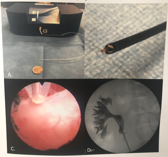
Figure 1: A, The Doppler transducer with a 20 Pulsed Doppler transceiver unit. B, Doppler transducer seen fully inserted into a ureteroscope. C, Doppler transducer seen in a ureteroscopic view while inside the porcine kidney. D, Retrograde pyelogram of the porcine ureter with ureteroscope inserted.
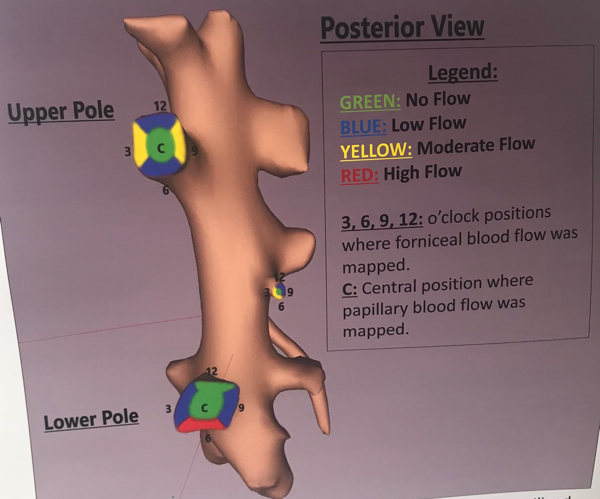
Figure 2: 3D reconstruction of the posterior calyces with blood flow outlined by color.
Presented by: Mitchell O’Leary, Department of Urology, University of California, Irvine
Co-Authors: Roshan M. Patel1, Kamaljot S. Kaler1, Mitchell O’Leary1, Vinay Cooper1, Egor Parkhomenko1, David Regan2, Jaime Landman1, and Ralph V. Clayman1
Author Affiliation: 1Department of Urology, University of California, Irvine, USA; 2Vascular Technology Incorporated, Nashua, USA
Written by: Zachary Valley Twitter: @ZacharyAValley, Department of Urology, University of California-Irvine, medical writer for UroToday.com at the 36th World Congress of Endourology (WCE) and SWL - September 20-23, 2018 Paris, France