A new technique introduced by Dr. Barentsz is nano-MRI, using ferrotran (ferumoxtran-10), which is a contrast agent that consists of nano-sized iron oxide particles.
At the cellular level, the region of the lymph node that does not have microscopic tumor consists of macrophages that uptake the iron particle, which appears black on MRI. Thus, metastases (no iron uptake in the lymph node) appear white on MRI and normal lymph node tissue is black. Presenting unpublished data from his center, Dr. Barentsz notes that compared with 68Ga-PSMA PET/CT, nano-MRI was able to more frequently detect positive lymph nodes that were <4 mm in size.
Based on recently published data suggesting that salvage lymph node dissection does not have long term benefit among patients after radical prostatectomy,1 new treatment modalities are needed to improve patient outcomes, particularly when these microscopic lymph nodes are detected. One such modality according to Dr. Barentsz is radiotherapy guided by ultra-small superparamagnetic iron oxide (USPIO)-contrast MRI staging. In this pioneering work, all patients received a boost of 60 Gy in 2-2.4 Gy/fraction or a sequential boost to 70.2 Gy in 1.8 Gy/fraction based on positive nano-MRI lymph nodes (all patients received >= 6 months of ADT). At the discretion of the physician, radiotherapy was given electively to lower chain lymph nodes when common iliac nodes were positive. With over 5 years of follow-up, as follows are the results:
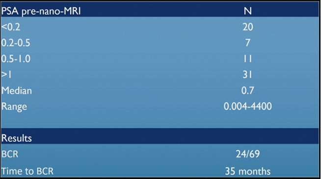
Based on their early experience, Dr. Barentsz states that for high-risk biochemical recurrence (BCR) patients, nano-MRI radiotherapy appears to be feasible and well tolerated with encouraging biochemical control rates. However, future multi-institutional prospective studies are needed to validate the clinical utility and efficacy of nano-MRI therapies.
Dr. Barentsz concluded with the following take-home messages:
- Most positive lymph nodes are small at <4mm
- Pelvic lymph node dissection and PSMA PET/CT have problems finding these small lymph nodes
- Early work using nano-MRI guided radiotherapy show promising results
Presented by: Jelle O. Barentsz, MD, PhD, Professor, Radboud University Medical Center, Nijmegen, The Netherlands
Written by: Zachary Klaassen, MD, MSc – Assistant Professor of Urology, Georgia Cancer Center, Augusta University/Medical College of Georgia, Twitter: @zklaassen_md at the 12th European Multidisciplinary Congress on Urological Cancers (EMUC) (#EMUC20 ), November 13th - 14th, 2020