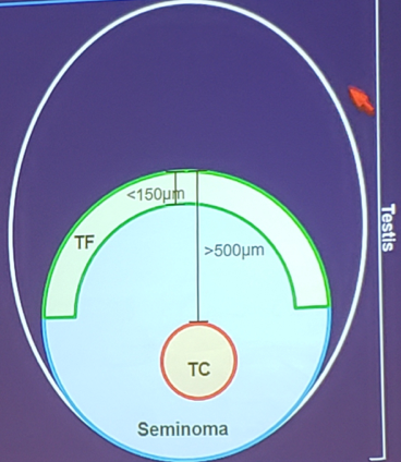
The hypothesis for this study was that different regional gene expression profiles are involved in molecular mechanisms leading to metastasis in testicular seminomas. For this study, the authors included formalin-fixed paraffin embedded (FFPE) tissue samples of 21 patients with clinical stage I disease, no adjuvant therapy and relapse-free survival of at least 2 years, and 14 patients showing metastasis (CSII/III). The tumor front and tumor center regions of each patient were determined and separately collected using laser capture microdissection. The depiction of tumor front and tumor center regions is as follows (TF – tumor front; TC – tumor center):
RNA was extracted and a multiplex gene expression analysis was performed on all tumor front and tumor center samples using nCounter technology of Nanostring. Furthermore, a panel of 770 transcripts was analyzed using the PanCancer Progression panel. Finally, different bioinformatics tools were employed for analyzing the expression data.
The authors found that differential gene expression patterns were observed in the metastatic and non-metastatic patients, with respect to both the tumor front and tumor center regions.
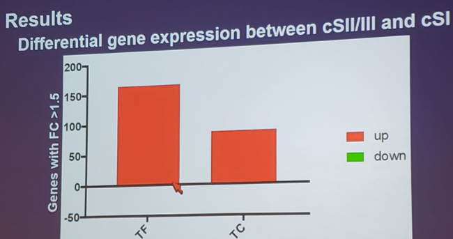
Ingenuity pathway analysis on the differentially expressed genes showed enrichment of tumor functions such as migration, invasion, and angiogenesis at the tumor front as compared to the tumor center. Interestingly, prominent inflammatory and cancer related pathways such as IL-6 signaling, acute phase response signaling, NF-κB signaling and, dendritic cell maturation were significantly upregulated in the metastatic versus non-metastatic tumors (p-value < 0.05).
The authors report, for the first time, tumor heterogeneity among patients with seminoma germ cell tumors. Dr. Nestler concluded with several important points:
- Seminomas are heterogeneous, which may be a reason for differences in metastatic potential
- IL-6 signaling in metastatic seminomas:
- May be a prognostic marker as has previously been demonstrated in colorectal, gastric, ovarian and breast cancer
- May be a therapeutic target as has previously been demonstrated in rheumatoid arthritis (tocilizumab) and Castleman’s disease (situximab)
Co-Authors: Friederike Haidl, Maike Wittersheim, Priya Dalvi, Pia Paffenholz, Svenja Wagener-Ryczek, David J. K. P. Pfister, Martin Hellmich, Reinhard Büttner, Margarete Odenthal, Axel Heidenreich; Department of Urology and Uro-Oncology, University Hospital of Cologne, Cologne, Germany; Institute of Pathology, University Hospital of Cologne, Cologne, Germany; Institute of Medical Statistics and Computational Biology, University of Cologne, Cologne, Germany
Written by: Zachary Klaassen, MD, MSc – Assistant Professor of Urology, Georgia Cancer Center, Augusta University/Medical College of Georgia, Twitter: @zklaassen_md at the 2019 American Society of Clinical Oncology Genitourinary Cancers Symposium, (ASCO GU) #GU19, February 14-16, 2019 - San Francisco, CA